
Local Anaesthetics
Learning Objectives
At the end of this chapter, the Phase II MBBS student will be able to:
- Classify local anaesthetics into esters and amides and explain the metabolic and allergenic difference. (PH1.17 — Knows)
- Describe the mechanism of action of local anaesthetics — sodium-channel blockade from the intracellular side with use-dependence. (PH1.17 — Knows)
- Explain how pKa determines onset and why local anaesthetics fail in infected tissue. (PH1.17 — Knows)
- Describe the individual agents — lidocaine, bupivacaine, ropivacaine, prilocaine and cocaine — with their characteristic properties. (PH1.17 — Knows)
- Explain the role of adrenaline and bicarbonate as adjuvants, including the digital-block caution. (PH1.17 — Knows)
- Describe the techniques of local anaesthesia. (PH1.17 — Knows)
- Recognise local anaesthetic systemic toxicity and manage it with lipid emulsion. (PH1.17 — Shows-how)
- State the maximum safe doses of lidocaine and bupivacaine. (PH1.17 — Knows)
- Explain why bupivacaine is the most cardiotoxic local anaesthetic. (PH1.17 — Knows)
Must-Know Summary
Local anaesthetics reversibly block nerve conduction without loss of consciousness, by blocking voltage-gated sodium channels — and everything about their clinical use follows from their structure. Esters (procaine, tetracaine, cocaine) are hydrolysed by plasma esterases to PABA and are allergenic; amides (lidocaine, bupivacaine, ropivacaine) are hepatic-metabolised and rarely allergic. The mechanism is precise: the unionised drug crosses the membrane, the ionised form binds the channel from inside in its open/inactivated state, producing use-dependent block. Two safety lessons dominate: adrenaline prolongs the block but is forbidden in digital blocks, and bupivacaine is the most cardiotoxic agent, with lipid emulsion the rescue in systemic toxicity.
In one line each:
- Esters — hydrolysed by plasma esterases, allergenic (PABA)
- Amides — hepatic metabolism, rarely allergic
- Local anaesthetics block — voltage-gated sodium channels from the intracellular side
- Use-dependent block — more nerve activity, more blockade
- pKa near physiological pH — faster onset
- Infected tissue is acidic — local anaesthetics fail there
- Adrenaline — prolongs the block, but never in a digital block
- Bupivacaine — the most cardiotoxic local anaesthetic
- Systemic toxicity — CNS first, then cardiovascular
- Lipid emulsion — the rescue for severe local anaesthetic toxicity
Classification
Box 1 — Local anaesthetics
- Esters (plasma-esterase hydrolysed, PABA, allergenic) — procaine, tetracaine, benzocaine, cocaine, chloroprocaine
- Amides (hepatic metabolism, rarely allergic) — lidocaine, bupivacaine, ropivacaine, levobupivacaine, prilocaine, mepivacaine
Box 2 — Techniques of local anaesthesia
- Surface/topical
- Infiltration
- Nerve block (plexus)
- Spinal (intrathecal)
- Epidural
- Intravenous regional (Bier's) block
Core Concepts
1. Classification and structure of local anaesthetics
A local anaesthetic consists of three parts: a lipophilic aromatic ring, an intermediate chain, and a hydrophilic amine. The nature of the intermediate chain divides the class into two groups with different fates:
- Esters — procaine, tetracaine (amethocaine), benzocaine, cocaine (and chloroprocaine). They are hydrolysed rapidly by plasma (pseudo)cholinesterases to para-aminobenzoic acid (PABA), which is the allergen responsible for ester hypersensitivity. They are therefore more allergenic and shorter-acting systemically. The mnemonic: esters have one "i" in their name (procaine, cocaine).
- Amides — lidocaine (lignocaine), bupivacaine, ropivacaine, prilocaine, mepivacaine (and levobupivacaine). They are metabolised in the liver (CYP-mediated N-dealkylation) and are rarely allergenic. The mnemonic: amides have two "i"s (lidocaine, bupivacaine).
This classification determines both the metabolism and the allergy profile — the first thing to establish about any local anaesthetic.
2. Mechanism — sodium channel blockade and use-dependence
Local anaesthetics reversibly block voltage-gated sodium channels, preventing the generation and conduction of action potentials in sensory (and motor) nerves. The mechanism has two steps that are frequently examined:
- Membrane penetration — the unionised (lipid-soluble) form crosses the axonal membrane.
- Channel blockade — inside the cell, the ionised (cationic) form binds the sodium channel in its open or inactivated state, blocking sodium influx.
Because the drug binds preferentially to the open/inactivated channel, the block is use-dependent (frequency-dependent) — the more the nerve fires, the more channels are blocked and the greater the block. The pKa determines the balance of unionised and ionised forms at physiological pH, and hence the onset: agents with a pKa close to physiological pH (7.4) — such as lidocaine (pKa ~7.9) — have more unionised drug available to penetrate and therefore a faster onset.
3. Differential nerve block and pH effects
Differential block. Small-diameter fibres are blocked before large ones, so the sequence of blockade is autonomic → pain and temperature → touch/proprioception → motor. This is why a regional block can produce analgesia with preserved motor function at low concentrations.
The pH effect and infected tissue. The ionisation of a local anaesthetic depends on the tissue pH. In acidic (infected) tissue, the increased hydrogen-ion concentration shifts the drug toward the ionised form, which cannot cross the membrane — so the anaesthetic fails to work in an abscess or inflamed tissue. This is the classic answer to "why does local anaesthesia fail in an infected site", and it also underlies the use of sodium bicarbonate to alkalinise the solution and hasten onset.
4. Individual agents
- Lidocaine (lignocaine) — the prototype amide: rapid onset, intermediate duration; used for infiltration, nerve blocks, topical anaesthesia, and (separately) as an antiarrhythmic (CH29).
- Bupivacaine — a long-acting, potent amide used for regional, spinal and epidural anaesthesia; it is the most cardiotoxic local anaesthetic (binds cardiac sodium channels avidly, causing ventricular arrhythmias and arrest that are difficult to resuscitate). Ropivacaine (its S-enantiomer) and levobupivacaine are the less cardiotoxic alternatives.
- Prilocaine — an amide metabolised to o-toluidine, which oxidises haemoglobin to methaemoglobinaemia — its characteristic toxicity (a risk with high doses or in infants).
- Cocaine — the only local anaesthetic that causes vasoconstriction (it blocks noradrenaline reuptake); used topically (ENT), but with abuse and cardiovascular risks.
- Tetracaine — a potent, long-acting ester used topically and for spinal anaesthesia.
- Benzocaine — a topical ester; rarely causes methaemoglobinaemia.
5. Adjuvants — adrenaline and bicarbonate
Adrenaline is added to local anaesthetic solutions to produce vasoconstriction, which prolongs the block, reduces bleeding and limits systemic absorption (and toxicity). It is contraindicated in digital (finger/toe/penis) blocks, where the vasoconstriction can cause ischaemic necrosis of the end-arterial tissue.
Sodium bicarbonate is added to raise the pH, increasing the unionised fraction and thereby hastening the onset of the block.
6. Techniques and systemic toxicity
Techniques. Local anaesthetics are administered by surface/topical application (skin, mucous membranes), infiltration (injection into the tissues), nerve block (around a nerve trunk or plexus), spinal (intrathecal) and epidural anaesthesia, and intravenous regional (Bier's) block.
Systemic toxicity results from excessive plasma concentrations (overdose or inadvertent intravascular injection). The clinical sequence is CNS first — circumoral tingling/numbness, metallic taste, tinnitus, light-headedness, then seizures — followed by cardiovascular depression: hypotension, bradycardia, arrhythmias and cardiac arrest (the cardiovascular toxicity being greatest with bupivacaine). Management is supportive (airway, oxygen, ventilation, benzodiazepines for seizures, cardiovascular support) with lipid emulsion (intralipid 20%) as the specific rescue for severe cardiotoxicity — the lipid "sink" binds the lipophilic anaesthetic. Maximum safe doses guide prevention: lidocaine ~3–4.5 mg/kg (up to 7 mg/kg with adrenaline) and bupivacaine ~2 mg/kg.
Tables
Table 1 — Esters versus amides
| Feature | Esters | Amides |
|---|---|---|
| Examples | Procaine, tetracaine, cocaine | Lidocaine, bupivacaine, ropivacaine |
| Metabolism | Plasma esterases → PABA | Hepatic |
| Allergy | More (PABA) | Rare |
| Mnemonic | One "i" | Two "i"s |
Table 2 — Individual local anaesthetics
| Agent | Class | Onset | Duration | Key point |
|---|---|---|---|---|
| Lidocaine | Amide | Rapid | Intermediate | Prototype, antiarrhythmic |
| Bupivacaine | Amide | Slow | Long | Most cardiotoxic |
| Ropivacaine | Amide | Slow | Long | Less cardiotoxic |
| Prilocaine | Amide | Rapid | Intermediate | Methaemoglobinaemia |
| Cocaine | Ester | — | — | Vasoconstriction |
| Tetracaine | Ester | Slow | Long | Potent, topical/spinal |
Table 3 — Adjuvants
| Adjuvant | Effect | Caution |
|---|---|---|
| Adrenaline | Vasoconstriction, prolongs block | Never in digital blocks |
| Sodium bicarbonate | Raises pH, faster onset | — |
Table 4 — Techniques of local anaesthesia
| Technique | Description |
|---|---|
| Surface/topical | Skin, mucous membranes |
| Infiltration | Into tissues |
| Nerve block | Around nerve trunk/plexus |
| Spinal | Intrathecal |
| Epidural | Epidural space |
Table 5 — Local anaesthetic systemic toxicity
| System | Features |
|---|---|
| CNS (first) | Circumoral tingling, tinnitus, seizures |
| Cardiovascular | Hypotension, arrhythmias, arrest |
| Management | Supportive + lipid emulsion |
| Seizures | Benzodiazepines |
Table 6 — Maximum safe doses
| Agent | Dose (no adrenaline) |
|---|---|
| Lidocaine | 3–4.5 mg/kg (7 mg/kg with adrenaline) |
| Bupivacaine | ~2 mg/kg |
Figures
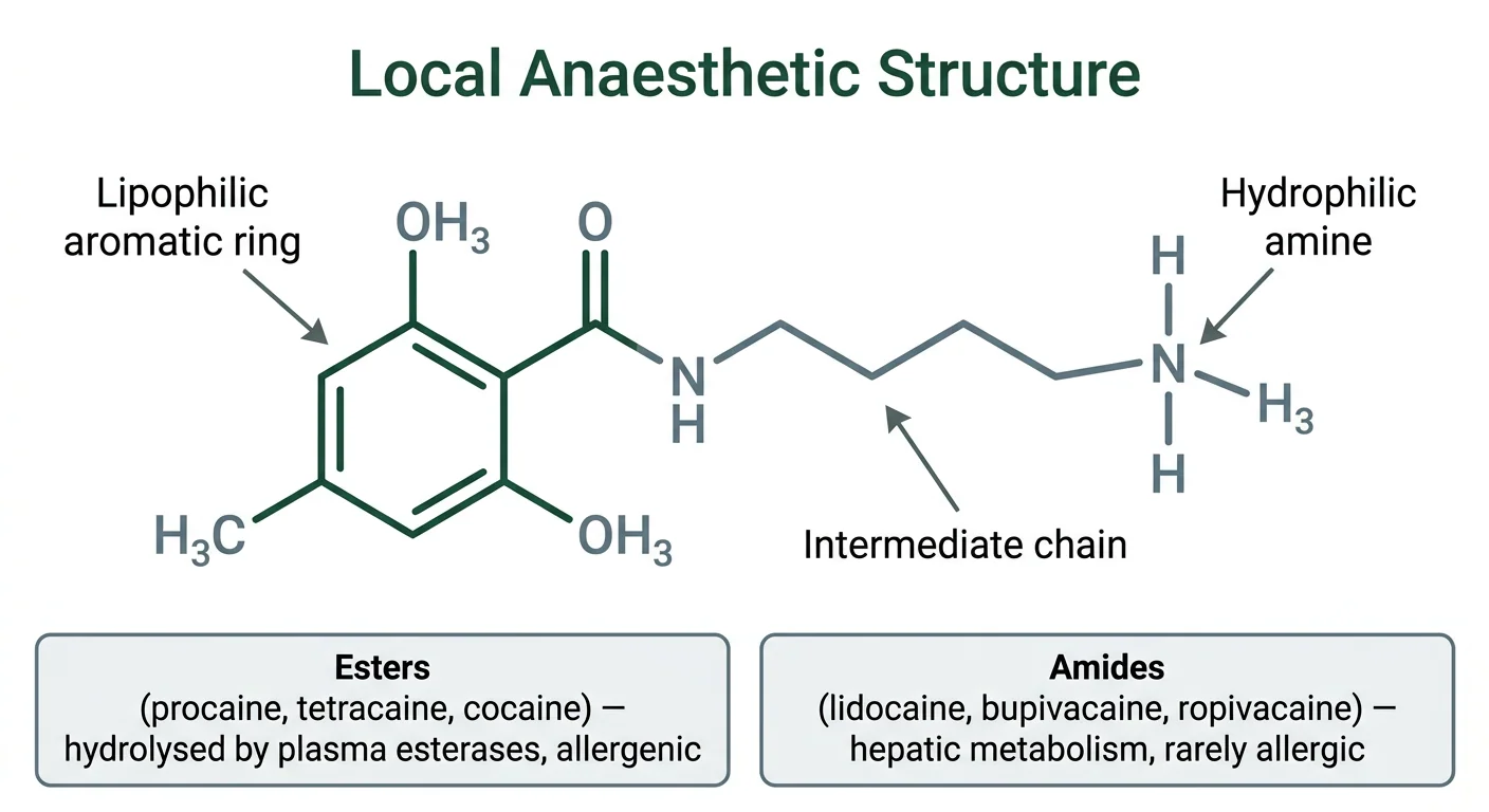
Figure 1 — Local anaesthetic structure and classification. Diagram of the three-part structure of a local anaesthetic — aromatic ring, intermediate chain and amine — with the ester and amide classes and their metabolic and allergic differences.
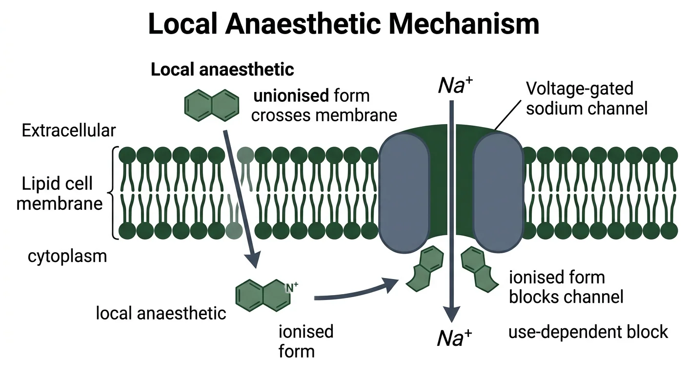
Figure 2 — Sodium channel blockade mechanism. Diagram of local anaesthetic action showing the unionised form crossing the nerve membrane and the ionised form binding the voltage-gated sodium channel from inside, producing use-dependent blockade.
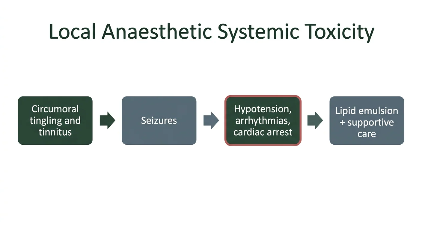
Figure 3 — Local anaesthetic systemic toxicity. Diagram of the progression of local anaesthetic systemic toxicity from circumoral tingling and tinnitus through seizures to cardiovascular collapse, with lipid emulsion and supportive care as treatment.
Clinical Correlation
Vignette 1 — Local anaesthetic systemic toxicity
During an epidural, a patient develops circumoral tingling, a metallic taste and tinnitus, then a generalised seizure.
Reasoning: This is local anaesthetic systemic toxicity — the CNS features (circumoral tingling, tinnitus, then seizures) appear first, before cardiovascular collapse. Management is immediate: stop the injection, secure the airway, give oxygen and a benzodiazepine for the seizure, and prepare for the cardiovascular consequences — with lipid emulsion (intralipid 20%) as the specific rescue for severe cardiotoxicity. Prevention is by adhering to maximum safe doses and aspirating before injection.
Vignette 2 — Bupivacaine cardiotoxicity
A patient inadvertently receives an intravascular dose of bupivacaine and develops refractory ventricular arrhythmias.
Reasoning: Bupivacaine is the most cardiotoxic local anaesthetic — it binds cardiac sodium channels avidly and dissociates slowly, causing ventricular arrhythmias and arrest that are difficult to resuscitate. This is why the less cardiotoxic alternatives ropivacaine and levobupivacaine are preferred where large doses are needed, and why lipid emulsion is the key rescue in bupivacaine-induced cardiac toxicity.
Vignette 3 — LA failure in an abscess
A surgeon attempts to incise a dental abscess after infiltration anaesthesia, but the patient still has full sensation.
Reasoning: In infected (acidic) tissue, the low pH shifts the local anaesthetic toward its ionised form, which cannot cross the nerve membrane — so the block fails. This is the classic explanation for local anaesthetic failure in an abscess, and it is why regional (nerve-block) anaesthesia away from the infected site, or alkalinisation, may be used instead.
Vignette 4 — Methaemoglobinaemia from prilocaine
A child given a large dose of prilocaine (as EMLA cream) develops cyanosis with a chocolate-brown blood colour that does not correct with oxygen.
Reasoning: Prilocaine is metabolised to o-toluidine, which oxidises haemoglobin to methaemoglobin — producing cyanosis and, in severe cases, tissue hypoxia. This is prilocaine's characteristic toxicity (a risk with high doses, in infants, or with the topical EMLA preparation). Management, when severe, is methylene blue, which reduces methaemoglobin back to haemoglobin.
Practical Linkage
Selecting the local anaesthetic
| Procedure | Agent | Maximum dose (adult) |
|---|---|---|
| Dental infiltration | Lidocaine + adrenaline | 7 mg/kg |
| Digital (finger) block | Lidocaine (no adrenaline) | 3–4.5 mg/kg |
| Spinal anaesthesia | Bupivacaine | ~2 mg/kg |
| Epidural (labour) | Ropivacaine | (less cardiotoxic) |
Exercise (PH1.17 — select the local anaesthetic and dose)
Discussion point
Why is adrenaline avoided in a digital block but useful in a dental block?
Expected: in a digital block the vasoconstriction risks ischaemic necrosis of the end-arterial digit, whereas in vascular dental tissue it usefully prolongs the block and reduces bleeding.
MCQ Bank
35 questions · tagged by topic, exam pattern & difficulty · full explanations
With regard to their mechanism of action, local anaesthetics produce their effect by:
Rapid Revision
- Local anaesthetics block — voltage-gated sodium channels
- Binding is intracellular — in the open or inactivated state
- Use-dependent block — more activity, more blockade
- pKa near physiological pH — faster onset
- Infected tissue is acidic — local anaesthetics fail
- Esters — plasma esterases, PABA, allergenic
- Amides — hepatic metabolism, rarely allergic
- Ester mnemonic — one "i"; amide — two "i"s
- Lidocaine — the prototype amide, also an antiarrhythmic
- Bupivacaine — the most cardiotoxic local anaesthetic
- Ropivacaine — the less cardiotoxic alternative
- Prilocaine — methaemoglobinaemia from o-toluidine
- Cocaine — the only local anaesthetic causing vasoconstriction
- Adrenaline — prolongs the block, but never in a digital block
- Bicarbonate — raises pH, hastens onset
- Systemic toxicity — CNS first, then cardiovascular
- Lipid emulsion — the rescue for severe cardiotoxicity
- Seizures in LA toxicity — benzodiazepines
- Lidocaine max dose — 3 to 4.5 mg/kg (7 with adrenaline)
- Bupivacaine max dose — about 2 mg/kg
- Differential block — autonomic, then pain, then motor
- Bier's block — intravenous regional anaesthesia
- EMLA cream — prilocaine, methaemoglobinaemia risk in infants
- Tetracaine — a long-acting ester for spinal anaesthesia
Viva Questions
- Classify local anaesthetics — Esters (procaine, tetracaine, cocaine) and amides (lidocaine, bupivacaine, ropivacaine).
- How do esters differ from amides — Esters are plasma-esterase hydrolysed to PABA and allergenic; amides are hepatic and rarely allergic.
- What is the mechanism of local anaesthetics — They block voltage-gated sodium channels from the intracellular side.
- What is use-dependent block — Blockade increases with nerve activity, as the drug binds the open channel.
- Why do local anaesthetics fail in infected tissue — The acidic pH increases the ionised fraction, which cannot cross the membrane.
- Why is adrenaline added — Vasoconstriction prolongs the block and reduces bleeding.
- Why is adrenaline avoided in digital blocks — Risk of ischaemic necrosis.
- Which is the most cardiotoxic local anaesthetic — Bupivacaine.
- What is the treatment of severe LA toxicity — Lipid emulsion, with supportive care.
- What causes prilocaine's cyanosis — Methaemoglobinaemia from o-toluidine, treated with methylene blue.
- What is the maximum dose of lidocaine — 3 to 4.5 mg/kg, up to 7 mg/kg with adrenaline.
- Why does cocaine cause vasoconstriction — It blocks noradrenaline reuptake.
- What is Bier's block — Intravenous regional anaesthesia.
- What is differential block — Small fibres (pain, autonomic) are blocked before large (motor) fibres.
References
- Tripathi KD. Essentials of Medical Pharmacology. 9th ed. New Delhi: Jaypee Brothers Medical Publishers; Chapter 26 (Local Anaesthetics).
- Katzung BG, Vanderah TW (eds). Basic & Clinical Pharmacology. 16th ed. New York: McGraw Hill; Chapter 26 (Local Anesthetics).
- Brunton LL, Knollmann BC (eds). Goodman & Gilman's The Pharmacological Basis of Therapeutics. 14th ed. New York: McGraw Hill; Chapter 24 (Local Anesthetics).
- Ritter JM, Flower RJ, Henderson G, et al. Rang & Dale's Pharmacology. 10th ed. Edinburgh: Elsevier; Chapter 46 (Local Anaesthetics).
- National Medical Commission. Competency Based Undergraduate Curriculum for the Indian Medical Graduate, Volume II — Pharmacology (Code: PH), competency PH1.17.
- Weinberg GL. Lipid emulsion infusion: resuscitation for local anesthetic and other drug overdose. Anesthesiology. 2012;117(1):180–187.
- Butterworth JF 4th, Strichartz GR. Molecular mechanisms of local anesthesia: a review. Anesthesiology. 1990;72(4):711–734.
- Becker DE, Reed KL. Essentials of local anesthetic pharmacology. Anesthesia Progress. 2006;53(3):98–108.
Downloads
All deliverables & printable revision sheets for CH19
Printable Chapter Handout & Study Notes
Generate a clean, high-yield PDF or print directly without sidebars and navigation menus.