
ANS Organisation, Neurotransmission & Cholinergic System
Learning Objectives
At the end of this chapter, the Phase II MBBS student will be able to:
- Describe the organisation of the autonomic nervous system and contrast the sympathetic and parasympathetic divisions anatomically and chemically. (PH1.14 — Knows)
- Explain the two-neuron relay and state why all preganglionic fibres are cholinergic, with the exceptions of the sweat glands, adrenal medulla and renal vessels. (PH1.14 — Knows)
- Describe the synthesis, storage, release and degradation of acetylcholine, naming the enzymes and the toxins that act at each step. (PH1.14 — Knows)
- Enumerate the muscarinic receptor subtypes M1–M5 with their locations, G-protein coupling and tissue effects. (PH1.14 — Knows)
- Differentiate nicotinic Nm from Nn receptors in location, structure and effect. (PH1.14 — Knows)
- Explain the signal transduction of muscarinic (G-protein coupled) versus nicotinic (ion-channel) receptors. (PH1.14 — Knows)
- Classify the cholinergic drugs into direct-acting, indirect-acting and blocking groups. (PH1.14 — Knows)
- Predict the organ effects of acetylcholine and atropine from the receptor distribution. (PH1.14 — Knows-how)
- Map the receptor subtype, the effect of ACh and the effect of atropine for the heart, iris, bronchi, gut, bladder, glands and skeletal muscle. (PH1.14 — Shows-how)
Must-Know Summary
The autonomic nervous system runs the body's involuntary functions through a two-neuron relay, and the dividing line between its two divisions is the neurotransmitter released postganglionically — noradrenaline in the sympathetic, acetylcholine in the parasympathetic — while every preganglionic fibre, in both divisions, is cholinergic. Acetylcholine is synthesised by choline acetyltransferase, stored in vesicles, released by calcium-dependent exocytosis, and destroyed within milliseconds by acetylcholinesterase. Its receptors are of two families: the muscarinic (G-protein coupled; M1–M5) and the nicotinic (ligand-gated ion channels; Nm at the muscle, Nn in ganglia). The muscarinic subtypes obey a simple rule — odd numbers (M1, M3, M5) couple to Gq and excite; even numbers (M2, M4) couple to Gi and inhibit.
In one line each:
- Both divisions use a two-neuron relay with a cholinergic nicotinic preganglionic synapse
- Sympathetic postganglionic transmitter — noradrenaline; parasympathetic — acetylcholine
- Sweat glands are sympathetic but muscarinic — the classic exception
- The adrenal medulla is a modified sympathetic ganglion releasing adrenaline
- ACh synthesis — choline plus acetyl-CoA by choline acetyltransferase
- ACh degradation — acetylcholinesterase, within milliseconds
- Botulinum toxin — blocks ACh release; latrotoxin — causes massive release
- M2 in the heart couples to Gi — bradycardia; M3 on glands and smooth muscle couples to Gq
- Nicotinic Nm — muscle; Nn — ganglia and adrenal medulla
Classification
Box 1 — Cholinergic (cholinomimetic) drugs
I. Direct-acting (act on the receptor)
- Choline esters — acetylcholine, methacholine, carbachol, bethanechol
- Alkaloids — pilocarpine, muscarine (muscarinic); nicotine, lobeline (nicotinic)
II. Indirect-acting (anticholinesterases)
- Reversible — physostigmine, neostigmine, pyridostigmine, edrophonium, donepezil, rivastigmine
- Irreversible — organophosphates (malathion, parathion, sarin), carbamates (carbaryl)
III. Cholinergic blockers
- Antimuscarinic — atropine, hyoscine, ipratropium
- Ganglion blockers — hexamethonium, trimethaphan
- Neuromuscular blockers — succinylcholine (depolarising); tubocurarine, vecuronium (non-depolarising)
Box 2 — Muscarinic and nicotinic receptor subtypes
- M1 — ganglia, gastric parietal cells, CNS — Gq (IP3/DAG) — gastric acid
- M2 — SA node, atria, AV node — Gi (↓cAMP, ↑K⁺) — bradycardia
- M3 — glands, smooth muscle (iris, bronchi, gut, bladder), endothelium — Gq — secretion, contraction, NO vasodilation
- M4/M5 — CNS — Gi/Gq
- Nm — neuromuscular junction — ion channel — muscle contraction
- Nn — autonomic ganglia, adrenal medulla, CNS — ion channel — ganglionic transmission
Core Concepts
1. Organisation of the autonomic nervous system
The autonomic nervous system (ANS) regulates the visceral, involuntary functions of the body — heart rate, blood pressure, secretions, smooth-muscle tone, glandular activity and pupil size. It operates through a two-neuron relay: a preganglionic neuron whose cell body lies in the central nervous system synapses with a postganglionic neuron in an autonomic ganglion, and the postganglionic fibre innervates the effector organ.
The sympathetic (thoracolumbar) division arises from the thoracic and upper lumbar cord (T1–L2/3). Its preganglionic fibres are short (synapsing in the nearby paravertebral or prevertebral ganglia) and its postganglionic fibres are long, so the sympathetic system delivers a diffuse response. The parasympathetic (craniosacral) division arises from the brainstem (cranial nerves III, VII, IX, X) and sacral cord (S2–S4). Its preganglionic fibres are long (synapsing in ganglia close to or within the target organ) and its postganglionic fibres are short, producing a discrete, localised response.
The chemical dividing line is the postganglionic neurotransmitter: sympathetic postganglionic fibres release noradrenaline (with adrenaline and dopamine as co-transmitters), while parasympathetic postganglionic fibres release acetylcholine. Crucially, all preganglionic fibres — of both divisions — release acetylcholine acting on nicotinic (Nn) receptors in the ganglion. Three exceptions recur in examinations: (1) the sympathetic fibres to sweat glands are cholinergic (muscarinic, M3) — sympathetic anatomically, cholinergic chemically; (2) the adrenal medulla is a modified sympathetic ganglion, innervated by a cholinergic preganglionic fibre and releasing adrenaline and noradrenaline into the blood; and (3) the sympathetic fibres to renal blood vessels are dopaminergic (D1).
2. Cholinergic neurotransmission — synthesis, storage, release and degradation
Synthesis. Acetylcholine is synthesised in the cholinergic nerve terminal from choline and acetyl-coenzyme A, catalysed by choline acetyltransferase (ChAT). Choline is taken up into the nerve terminal by a sodium-dependent choline transporter — the rate-limiting step — and acetyl-CoA is supplied by mitochondria. The uptake of choline is blocked by hemicholinium, which therefore limits ACh synthesis.
Storage. ACh is packaged into synaptic vesicles by the vesicular ACh transporter; vesamicol blocks this transport and depletes vesicular ACh.
Release. Arrival of the action potential opens voltage-gated calcium channels; the rise in intracellular Ca²⁺ triggers fusion of vesicles with the presynaptic membrane and exocytosis of ACh. Botulinum toxin cleaves SNARE proteins essential for this fusion and thereby blocks ACh release, producing flaccid paralysis; black widow spider venom (alpha-latrotoxin) causes massive, uncontrolled release of ACh, depleting the store.
Degradation. Released ACh is hydrolysed within milliseconds by acetylcholinesterase (AChE), an enzyme concentrated at the synapse, into choline and acetate; the choline is taken back up for resynthesis. AChE has two functional sites — an anionic site (binds the quaternary nitrogen) and an esteratic site (binds and hydrolyses the ester) — the structural basis of anticholinesterase drug action (CH10). A related but non-specific enzyme, butyrylcholinesterase (plasma cholinesterase, pseudocholinesterase), hydrolyses ACh and other esters in the plasma (its deficiency causes succinylcholine apnoea — CH07).
3. Muscarinic receptor subtypes and signal transduction
Muscarinic receptors are G-protein coupled (metabotropic) receptors activated by muscarine and blocked by atropine. Five subtypes (M1–M5) exist, and their locations, coupling and effects are the single most reproduced table in the autonomic block.
- M1 — "neural": autonomic ganglia, CNS, and gastric parietal cells (via the vagus). Coupling Gq → phospholipase C → IP3/DAG → ↑Ca²⁺. Mediates gastric acid secretion and CNS excitation.
- M2 — "cardiac": SA node, atria and AV node. Coupling Gi → ↓adenylyl cyclase (↓cAMP), and ↑K⁺ conductance → bradycardia, slowed AV conduction. This is why atropine (which blocks M2) causes tachycardia.
- M3 — "glandular/smooth muscle": exocrine glands, and smooth muscle of the iris, bronchi, gut and bladder. Coupling Gq → contraction and secretion; on the vascular endothelium M3 releases nitric oxide, producing vasodilation.
- M4/M5 — predominantly in the CNS (M4 couples Gi, M5 Gq); rarely tested individually.
The mnemonic is "odd = Gq (excitatory), even = Gi (inhibitory)" — M1, M3 and M5 couple to Gq; M2 and M4 to Gi. The tissue effects of muscarinic stimulation are therefore: bradycardia (M2), miosis and accommodation (M3), bronchoconstriction and increased secretions (M3), increased gut motility and bladder contraction (M3), salivation, lacrimation and sweating (M3), and vasodilation via endothelial NO (M3).
4. Nicotinic receptors and the pharmacology of ACh
Nicotinic receptors are ligand-gated ion channels (ionotropic) — pentameric channels that open on agonist binding, permitting Na⁺ influx and rapid depolarisation. They are of two principal subtypes:
- Nm (muscle) — at the neuromuscular junction of skeletal muscle, mediating muscle contraction (blocked by neuromuscular blockers, CH11).
- Nn (neuronal) — in the autonomic ganglia, the adrenal medulla and the CNS, mediating ganglionic transmission (blocked by ganglion blockers).
A key pharmacological property of nicotinic receptors is depolarisation block: a persistent agonist (nicotine, or the depolarising blocker succinylcholine) first stimulates, then — because the channel stays depolarised and the membrane becomes refractory — produces a blockade. This distinction between stimulation and depolarisation block underlies the muscle relaxant pharmacology of CH11.
Actions of acetylcholine itself are the sum of muscarinic and nicotinic effects — the "muscarinic" effects (bradycardia, secretions, smooth-muscle contraction, miosis, vasodilation) are those reproduced by muscarine and blocked by atropine; the "nicotinic" effects (ganglionic stimulation, skeletal-muscle contraction) are those reproduced by nicotine. Acetylcholine is clinically useless as a systemic drug because it is destroyed by cholinesterases within milliseconds.
5. Classification of cholinergic drugs
Cholinergic (cholinomimetic) drugs either mimic or enhance the actions of acetylcholine; cholinergic blockers oppose them.
Cholinomimetics — direct-acting (act on the receptor):
- Choline esters: acetylcholine, methacholine, carbachol, bethanechol.
- Alkaloids: pilocarpine, muscarine (muscarinic); nicotine (nicotinic).
Cholinomimetics — indirect-acting (anticholinesterases) (increase ACh by inhibiting its breakdown):
- Reversible: physostigmine, neostigmine, pyridostigmine, edrophonium, donepezil, rivastigmine, galantamine.
- Irreversible: organophosphates (malathion, parathion, sarin), carbamates (carbaryl).
Cholinergic blockers:
- Antimuscarinic: atropine, hyoscine, ipratropium, and their congeners.
- Antinicotinic: ganglion blockers (rarely used) and neuromuscular blockers (depolarising — succinylcholine; non-depolarising — tubocurarine, vecuronium, atracurium, rocuronium).
The direct and indirect agonists, and the anticholinesterases, are developed drug-by-drug in CH10; the antimuscarinics and neuromuscular blockers in CH11.
Tables
Table 1 — Sympathetic versus parasympathetic
| Feature | Sympathetic | Parasympathetic |
|---|---|---|
| Origin | Thoracolumbar (T1–L2/3) | Craniosacral (CN III, VII, IX, X; S2–S4) |
| Preganglionic fibre | Short | Long |
| Postganglionic fibre | Long | Short |
| Postganglionic transmitter | Noradrenaline | Acetylcholine |
| Response pattern | Diffuse | Discrete |
| Ganglion type | Paravertebral/prevertebral | Near or in the target organ |
Table 2 — Muscarinic receptor subtypes
| Subtype | Location | G-protein | Second messenger | Effect |
|---|---|---|---|---|
| M1 | Ganglia, parietal cells, CNS | Gq | IP3/DAG, ↑Ca²⁺ | Gastric acid, CNS excitation |
| M2 | SA node, atria, AV node | Gi | ↓cAMP, ↑K⁺ | Bradycardia, ↓ conduction |
| M3 | Glands, smooth muscle, endothelium | Gq | IP3/DAG, ↑Ca²⁺ | Secretion, contraction, NO vasodilation |
| M4 | CNS | Gi | ↓cAMP | CNS |
| M5 | CNS | Gq | IP3/DAG | CNS |
Table 3 — Nicotinic receptor subtypes
| Feature | Nm (muscle) | Nn (neuronal) |
|---|---|---|
| Location | Neuromuscular junction | Autonomic ganglia, adrenal medulla, CNS |
| Structure | Ligand-gated ion channel | Ligand-gated ion channel |
| Effect | Skeletal muscle contraction | Ganglionic transmission |
| Prototype blocker | Neuromuscular blockers | Ganglion blockers |
Table 4 — Steps of cholinergic neurotransmission
| Step | Process | Enzyme / toxin |
|---|---|---|
| 1 | Choline uptake | Hemicholinium blocks |
| 2 | Synthesis | Choline acetyltransferase (ChAT) |
| 3 | Vesicular storage | Vesamicol blocks |
| 4 | Release | Botulinum blocks; latrotoxin causes release |
| 5 | Degradation | Acetylcholinesterase |
Table 5 — Toxins acting on the cholinergic synapse
| Toxin | Site | Effect |
|---|---|---|
| Hemicholinium | Choline transporter | Depletes ACh synthesis |
| Vesamicol | Vesicular transporter | Depletes stored ACh |
| Botulinum toxin | SNARE proteins | Blocks ACh release |
| Black widow venom (latrotoxin) | Presynaptic terminal | Massive ACh release |
| Organophosphates | Acetylcholinesterase | ACh accumulation |
Table 6 — Cholinergic drug classification
| Group | Mechanism | Examples |
|---|---|---|
| Direct choline esters | Receptor agonist | Bethanechol, carbachol |
| Direct alkaloids | Receptor agonist | Pilocarpine, nicotine |
| Reversible anticholinesterases | Inhibit AChE | Neostigmine, physostigmine |
| Irreversible anticholinesterases | Inhibit AChE | Organophosphates |
| Antimuscarinic | Block M receptors | Atropine, ipratropium |
| Neuromuscular blockers | Block Nm | Succinylcholine, vecuronium |
Figures
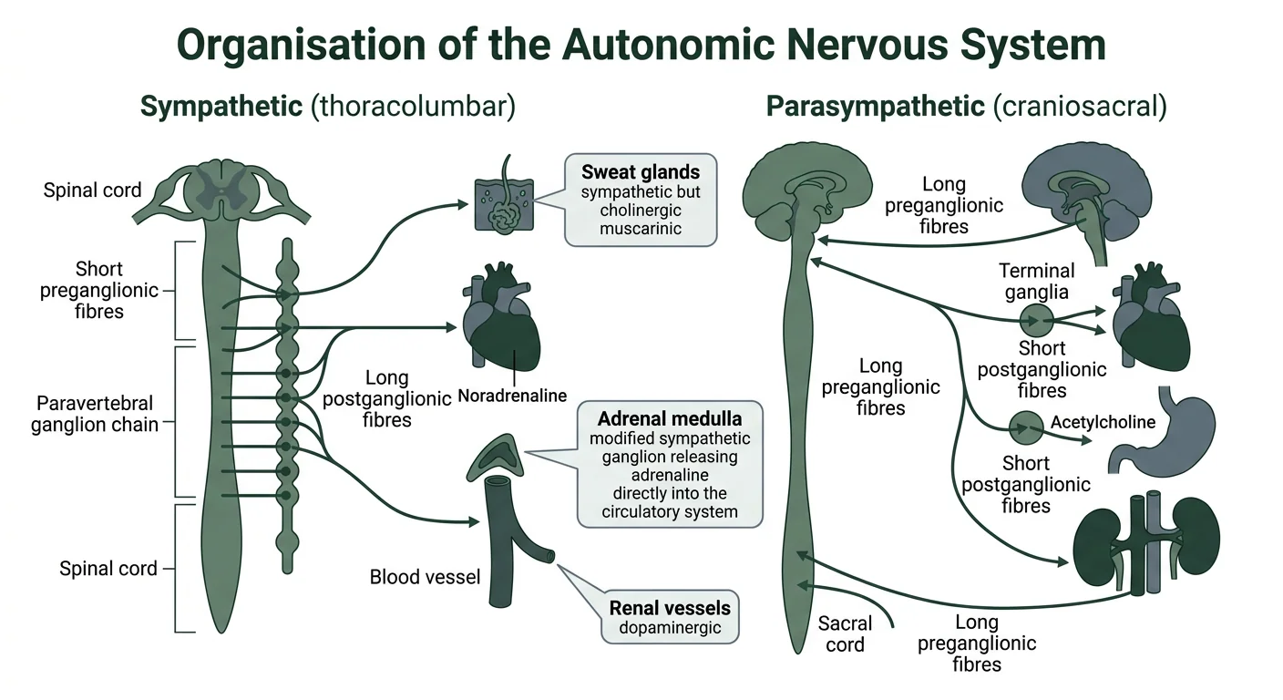
Figure 1 — Organisation of the autonomic nervous system. Diagram contrasting the sympathetic thoracolumbar and parasympathetic craniosacral divisions of the autonomic nervous system, showing the two-neuron relay, the noradrenaline versus acetylcholine postganglionic transmitters, and the exceptions of sweat glands, adrenal medulla and renal vessels.
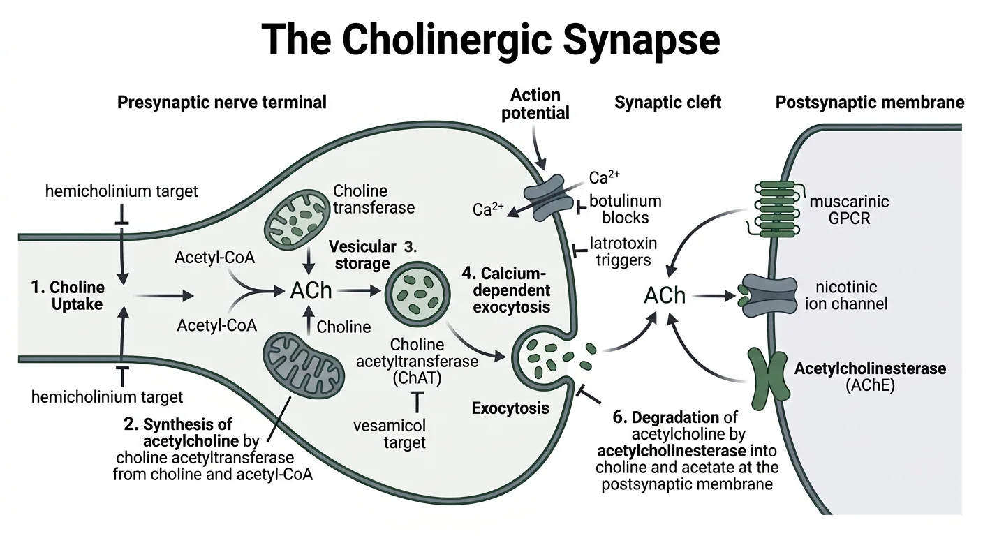
Figure 2 — The cholinergic synapse. Diagram of the cholinergic synapse showing synthesis, storage, release and degradation of acetylcholine, with the enzyme and toxin targets at each step including choline acetyltransferase, acetylcholinesterase, hemicholinium, vesamicol, botulinum toxin and latrotoxin.
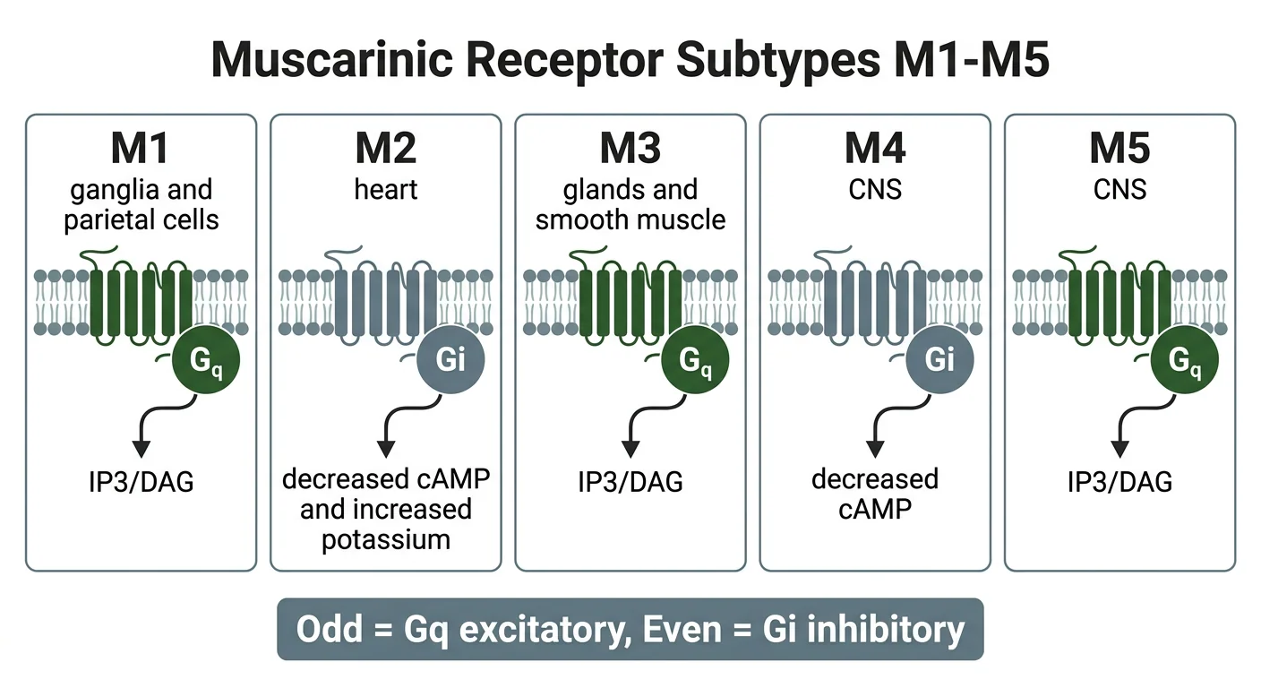
Figure 3 — Muscarinic receptor subtypes and signal transduction. Diagram of the five muscarinic receptor subtypes M1 through M5 with their locations and G-protein coupling, showing odd-numbered receptors coupling to Gq and even-numbered to Gi, and the resulting second messengers.
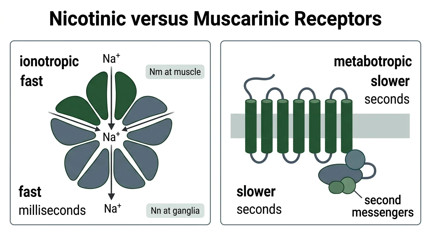
Figure 4 — Nicotinic versus muscarinic receptor structure. Comparison of the nicotinic receptor as a pentameric ligand-gated ion channel permitting sodium influx, against the muscarinic receptor as a seven-transmembrane G-protein coupled receptor acting through second messengers.
Clinical Correlation
Vignette 1 — The organophosphate toxidrome
A farmer who sprayed insecticide without protection is brought in salivating, sweating, with pin-point pupils, bronchial secretions, abdominal cramps, diarrhoea and muscle fasciculations, with a heart rate of 48/minute.
Reasoning: Organophosphates inhibit acetylcholinesterase, so acetylcholine accumulates at every cholinergic synapse. The muscarinic effects explain the salivation, lacrimation, bronchial secretions, miosis, bradycardia (M2), cramps and diarrhoea (M3) — the "SLUDGE" toxidrome — while the nicotinic effects at the neuromuscular junction (Nm) explain the fasciculations and, later, weakness. The vignette shows why the toxidrome is the direct expression of the receptor map taught in this chapter: every sign can be traced to a specific receptor subtype, which is also the logic of treating the muscarinic signs with atropine (M-blockade) and regenerating the enzyme with pralidoxime (developed in CH10).
Vignette 2 — Botulism
A patient who ate home-canned food develops blurred vision, dry mouth, and a descending flaccid paralysis with normal sensation.
Reasoning: Botulinum toxin cleaves the SNARE proteins required for vesicle fusion, so acetylcholine release is blocked at the neuromuscular junction (Nm) and autonomic cholinergic synapses. The result is flaccid paralysis with anticholinergic features (dry mouth, dilated pupils, ileus) — the mirror image of organophosphate poisoning. The contrast with myasthenia gravis (an immune attack on the receptor rather than a toxin blocking release) is a classic discriminator: both cause weakness, but botulism also blocks autonomic cholinergic transmission.
Vignette 3 — Atropine premedication
Before a general anaesthetic, a patient receives intramuscular atropine and develops a dry mouth, a heart rate of 100/minute, and dilated pupils.
Reasoning: Atropine blocks muscarinic receptors, so its effects are exactly the inverse of muscarinic stimulation: M3 blockade dries secretions (mouth, bronchi) and dilates the pupil (relaxing the iris sphincter), while M2 blockade removes vagal tone and produces tachycardia. These effects are used deliberately before anaesthesia — to reduce secretions and prevent vagal bradycardia during intubation — and the triad of dry mouth, tachycardia and mydriasis is the bedside signature of muscarinic blockade, developed fully in CH11.
Vignette 4 — Fasciculations then paralysis
During induction, a patient given succinylcholine first shows brief, coarse muscle fasciculations, followed by flaccid paralysis.
Reasoning: Succinylcholine is a nicotinic agonist at the Nm receptor. It initially opens the ion channel and depolarises the muscle, causing visible fasciculations; but because it is not rapidly hydrolysed like ACh, the membrane remains depolarised and refractory, so subsequent impulses cannot propagate — a depolarisation block. This sequence of stimulation-then-paralysis is the defining feature of depolarising neuromuscular blockade, and it is why the drug is preceded by a small dose of a non-depolarising blocker in some protocols (to blunt the fasciculations).
Practical Linkage
Receptor-distribution map
| Organ | Receptor | Effect of ACh | Effect of atropine |
|---|---|---|---|
| SA node (heart) | M2 | Bradycardia | Tachycardia |
| Iris (sphincter pupillae) | M3 | Miosis | Mydriasis |
| Bronchi | M3 | Bronchoconstriction, secretion | Bronchodilation, dryness |
| Gut | M3 | Increased motility | Reduced motility |
| Bladder (detrusor) | M3 | Contraction | Relaxation, retention |
| Salivary glands | M3 | Salivation | Dry mouth |
| Skeletal muscle | Nm | Contraction | No effect (nicotinic) |
| Autonomic ganglia | Nn | Transmission | No effect (atropine is muscarinic) |
Exercise (PH1.14 — map the receptor distribution)
Complete the table for each organ: the receptor subtype, the effect of acetylcholine, and the effect of atropine.
Discussion point
Why does atropine not block skeletal muscle contraction?
Expected: atropine is a muscarinic antagonist and does not act on the nicotinic Nm receptor; muscle blockade requires a neuromuscular blocker such as tubocurarine or succinylcholine.
MCQ Bank
35 questions · tagged by topic, exam pattern & difficulty · full explanations
The postganglionic neurotransmitter of the parasympathetic nervous system is:
Rapid Revision
- Both ANS divisions use a two-neuron relay with a cholinergic nicotinic preganglionic synapse
- Sympathetic postganglionic transmitter — noradrenaline
- Parasympathetic postganglionic transmitter — acetylcholine
- Sweat glands — sympathetic but cholinergic muscarinic
- Adrenal medulla — a modified sympathetic ganglion releasing adrenaline
- Renal sympathetic fibres — dopaminergic, D1
- ACh synthesis — choline plus acetyl-CoA, by choline acetyltransferase
- Rate-limiting step in ACh synthesis — choline uptake
- ACh degradation — acetylcholinesterase, within milliseconds
- AChE has two sites — anionic and esteratic
- Hemicholinium — blocks choline uptake
- Vesamicol — blocks vesicular storage
- Botulinum toxin — blocks ACh release
- Latrotoxin — causes massive ACh release
- M1 — ganglia and parietal cells, Gq, gastric acid
- M2 — SA node, Gi, bradycardia
- M3 — glands and smooth muscle, Gq, secretion and contraction
- Odd muscarinic receptors — Gq; even — Gi
- M3 on endothelium — releases nitric oxide, vasodilation
- Nm — neuromuscular junction; Nn — ganglia and adrenal medulla
- Nicotinic receptors — pentameric ligand-gated ion channels
- Muscarinic receptors — G-protein coupled, metabotropic
- Persistent nicotinic agonist — depolarisation block
- Acetylcholine itself — clinically useless, destroyed in milliseconds
- Direct cholinomimetics — choline esters and alkaloids
- Indirect cholinomimetics — reversible and irreversible anticholinesterases
Viva Questions
- What is the postganglionic transmitter of the parasympathetic system — Acetylcholine, acting on muscarinic receptors.
- Why are all preganglionic fibres cholinergic — Both divisions release acetylcholine at the nicotinic (Nn) ganglion synapse; the divisions differ only in the postganglionic transmitter.
- Give the three autonomic exceptions — Sweat glands (sympathetic but muscarinic), adrenal medulla (modified ganglion releasing adrenaline), renal vessels (dopaminergic D1).
- Which enzyme synthesises acetylcholine — Choline acetyltransferase, from choline and acetyl-CoA.
- Which enzyme degrades acetylcholine — Acetylcholinesterase, within milliseconds, into choline and acetate.
- How does botulinum toxin cause paralysis — By cleaving SNARE proteins and blocking acetylcholine release.
- Where is the M2 receptor and what does it do — On the SA node, coupling to Gi, producing bradycardia.
- Where is the M3 receptor — On glands and smooth muscle (iris, bronchi, gut, bladder), producing secretion and contraction.
- What is the mnemonic for muscarinic coupling — Odd receptors (M1, M3, M5) couple to Gq; even (M2, M4) couple to Gi.
- How does muscarinic stimulation cause vasodilation — M3 receptors on the endothelium release nitric oxide.
- Differentiate Nm from Nn — Nm is the muscle nicotinic receptor at the neuromuscular junction; Nn is the neuronal receptor in ganglia and adrenal medulla.
- What is depolarisation block — Persistent nicotinic agonist action that first stimulates then blocks, as the membrane remains depolarised and refractory.
- Classify the cholinergic drugs — Direct-acting (choline esters, alkaloids), indirect-acting (anticholinesterases), and blockers (antimuscarinic, antinicotinic).
References
- Tripathi KD. Essentials of Medical Pharmacology. 9th ed. New Delhi: Jaypee Brothers Medical Publishers; Chapters 8 and 9 (Autonomic Nervous System; Cholinergic System).
- Katzung BG, Vanderah TW (eds). Basic & Clinical Pharmacology. 16th ed. New York: McGraw Hill; Chapters 6–8 (Cholinoceptor-Activating and Cholinesterase-Inhibiting Drugs).
- Brunton LL, Knollmann BC (eds). Goodman & Gilman's The Pharmacological Basis of Therapeutics. 14th ed. New York: McGraw Hill; Chapters 8 and 9 (Muscarinic Receptor Agonists and Antagonists).
- Ritter JM, Flower RJ, Henderson G, et al. Rang & Dale's Pharmacology. 10th ed. Edinburgh: Elsevier; Chapters 12 and 13 (Cholinergic Transmission).
- National Medical Commission. Competency Based Undergraduate Curriculum for the Indian Medical Graduate, Volume II — Pharmacology (Code: PH), competency PH1.14.
- Caulfield MP, Birdsall NJM. International Union of Pharmacology. XVII. Classification of muscarinic acetylcholine receptors. Pharmacological Reviews. 1998;50(2):279–290.
- Wessler I, Kirkpatrick CJ. Acetylcholine beyond neurons: the non-neuronal cholinergic system. British Journal of Pharmacology. 2008;154(8):1558–1571.
- Rang HP, Dale MM, Ritter JM, et al. Pharmacology. 7th ed. Edinburgh: Churchill Livingstone; section on the autonomic nervous system.
Downloads
All deliverables & printable revision sheets for CH09
Printable Chapter Handout & Study Notes
Generate a clean, high-yield PDF or print directly without sidebars and navigation menus.