
Pharmacodynamics — Receptors, Signal Transduction, Dose-Response
Learning Objectives
At the end of this chapter, the Phase II MBBS student will be able to:
- Define pharmacodynamics and enumerate the general mechanisms by which drugs produce their effects. (PH1.5 — Knows)
- Describe the non-receptor mechanisms of drug action with representative examples. (PH1.5 — Knows)
- Define a receptor and distinguish affinity from intrinsic activity. (PH1.5 — Knows)
- Classify receptors into the four superfamilies and compare them by structure, timescale and mode of transduction. (PH1.5 — Knows-how)
- Describe the G-protein cycle and relate Gs, Gi and Gq to their second messengers and representative receptors. (PH1.5 — Knows-how)
- Differentiate full agonists, partial agonists, inverse agonists and antagonists in terms of intrinsic activity. (PH1.5 — Knows-how)
- Construct and interpret graded and quantal dose–response curves and derive Emax, EC₅₀, ED₅₀, TD₅₀ and LD₅₀. (PH1.5 — Shows-how)
- Predict the effect of competitive, non-competitive and irreversible antagonists on the agonist dose–response curve. (PH1.5 — Shows-how)
- Calculate and interpret the therapeutic index and certain safety factor, and identify drugs with a narrow therapeutic index. (PH1.5 — Shows-how)
- Explain receptor regulation, desensitisation, tachyphylaxis and rebound phenomena, and relate them to clinical practice. (PH1.5 — Knows-how)
Must-Know Summary
Pharmacodynamics is what the drug does to the body. Where pharmacokinetics determined how much drug reaches the site of action, pharmacodynamics determines what happens when it arrives. Almost every drug acts by binding to a specific macromolecular target — usually a receptor, sometimes an enzyme, ion channel or transporter — and the character of that interaction explains both the therapeutic effect and the adverse one.
Two skills carry most of the examination weight. The first is reading a dose–response curve: recognising at sight whether a drug is a full agonist, a partial agonist, a competitive antagonist or a non-competitive antagonist. The second is knowing the receptor families and their second messengers, because that knowledge is reused in every systemic chapter that follows.
In one line each:
- Drugs do not confer new functions; they modulate existing physiological processes.
- Affinity is the tendency to bind; intrinsic activity is the ability to activate once bound.
- Agonist has affinity and intrinsic activity of 1; antagonist has affinity but intrinsic activity of 0; partial agonist lies between; inverse agonist has negative intrinsic activity.
- The four receptor superfamilies, in ascending order of timescale, are ligand-gated ion channels (milliseconds), G-protein coupled receptors (seconds), kinase-linked receptors (minutes) and nuclear receptors (hours).
- Gs raises cAMP, Gi lowers cAMP, Gq generates IP₃ and DAG and raises intracellular calcium.
- Cholera toxin irreversibly activates Gs; pertussis toxin inhibits Gi.
- Potency is the position of the curve on the dose axis; efficacy is the height of its plateau. Efficacy matters more clinically.
- A competitive antagonist shifts the curve parallel to the right with Emax unchanged and is surmountable.
- A non-competitive or irreversible antagonist lowers Emax and is insurmountable.
- A partial agonist has a lower Emax than a full agonist but may be more potent, and behaves as an antagonist in the presence of a full agonist.
- Graded curves measure intensity in one individual; quantal curves measure the proportion responding in a population.
- Therapeutic index = LD₅₀ / ED₅₀; the larger the value, the safer the drug.
- Tachyphylaxis is rapid tolerance developing over minutes to hours.
Classification
Box 1 — Mechanisms of drug action
I. Non-receptor mediated
- Physical action — bulk laxatives, activated charcoal, osmotic diuretics, radioactive isotopes
- Chemical action — antacids, chelating agents, protamine neutralising heparin
- Enzyme inhibition — aspirin, ACE inhibitors, statins, allopurinol, neostigmine, omeprazole
- Enzyme activation or reactivation — pralidoxime, phenobarbitone inducing UGT
- False substrate / antimetabolite — methyldopa, 5-fluorouracil, sulfonamides
- Action on ion channels — local anaesthetics, calcium channel blockers, sulfonylureas
- Action on transporters — SSRIs, digoxin on Na⁺/K⁺-ATPase, proton pump inhibitors, SGLT2 inhibitors
II. Receptor mediated
- Agonists — full, partial, inverse
- Antagonists — competitive, non-competitive, irreversible
- Allosteric modulators — positive (benzodiazepines) and negative
Box 2 — The four receptor superfamilies
- Type 1 — Ligand-gated ion channels (ionotropic) · timescale milliseconds — Nicotinic acetylcholine, GABA-A, glycine, NMDA and AMPA, 5-HT₃
- Type 2 — G-protein coupled receptors (metabotropic, 7-transmembrane) · timescale seconds
- Gs — increases cAMP: β₁, β₂, D₁, H₂, V₂
- Gi — decreases cAMP: α₂, D₂, M₂, opioid
- Gq — IP₃ and DAG, raises calcium: α₁, M₁, M₃, H₁, V₁, 5-HT₂
- Type 3 — Enzyme-linked (kinase-linked) receptors · timescale minutes
- Receptor tyrosine kinases — insulin, growth factors
- Cytokine receptors — JAK-STAT pathway
- Guanylyl cyclase — atrial natriuretic peptide; soluble form for nitric oxide
- Type 4 — Nuclear (intracellular) receptors · timescale hours
- Corticosteroids, sex steroids, thyroid hormone, vitamin D, retinoids
Core Concepts
1. Principles and non-receptor mechanisms of drug action
Pharmacodynamics is the study of the biochemical and physiological effects of drugs and of their mechanisms of action. The cardinal principle, worth restating from CH01, is that a drug cannot confer a new function on a tissue. It can only modulate the rate or extent of a process the tissue already performs, and it does so in one of four broad ways: stimulation (adrenaline on the heart), depression (quinidine on cardiac conduction), irritation (counter-irritants, and at higher intensity, tissue damage), or replacement and cytotoxic action (insulin in diabetes; anticancer and antimicrobial agents).
Most drugs act on one of four classes of regulatory macromolecule: receptors, enzymes, ion channels and transporters. A minority act without any specific macromolecular target at all.
Non-receptor mechanisms deserve early emphasis because students tend to assume all drug action is receptor-mediated.
- Physical action — the drug acts by a bulk physical property. Bulk laxatives such as ispaghula act by adsorbing water and increasing faecal mass; activated charcoal adsorbs toxins; mannitol acts by osmotic pressure; dimethicone alters surface tension; and the physical radioactivity of ¹³¹I destroys thyroid tissue.
- Chemical action — a simple chemical reaction. Antacids neutralise gastric acid; chelating agents such as desferrioxamine, penicillamine and dimercaprol bind heavy metals; protamine, a strongly basic protein, neutralises the strongly acidic heparin; oxidising antiseptics act chemically on microbial proteins.
- Enzyme inhibition — quantitatively one of the most important mechanisms in therapeutics. Aspirin irreversibly acetylates cyclo-oxygenase; ACE inhibitors block angiotensin converting enzyme; statins competitively inhibit HMG-CoA reductase; allopurinol inhibits xanthine oxidase; neostigmine inhibits acetylcholinesterase; omeprazole irreversibly inhibits the gastric H⁺/K⁺-ATPase; methotrexate inhibits dihydrofolate reductase.
- Enzyme activation or induction — phenobarbitone inducing glucuronyl transferase (CH03); pralidoxime reactivating phosphorylated cholinesterase.
- False substrate / antimetabolite action — the drug is accepted by an enzyme in place of the natural substrate and generates a useless or less active product. Methyldopa is converted to the false transmitter α-methylnoradrenaline; 5-fluorouracil substitutes for uracil; sulfonamides are false substrates for the folate synthesis pathway.
- Action on ion channels — local anaesthetics block voltage-gated sodium channels; calcium channel blockers block L-type channels; sulfonylureas block the ATP-sensitive potassium channel of the β cell; amiodarone blocks potassium channels.
- Action on transporters — SSRIs block the serotonin reuptake transporter; tricyclic antidepressants block noradrenaline and serotonin reuptake; digoxin inhibits the Na⁺/K⁺-ATPase; proton pump inhibitors act on the gastric proton pump; SGLT2 inhibitors block renal glucose reabsorption; probenecid blocks the organic anion transporter (CH03).
2. Receptors: definition, nature and regulation
A receptor is a specific macromolecule, usually a protein, with which a drug or endogenous ligand interacts to produce a characteristic biological response. The concept originated with Langley's "receptive substance" and Ehrlich's dictum that corpora non agunt nisi fixata — a substance does not act unless it is bound.
Two independent properties determine how a drug behaves at a receptor:
- Affinity — the tendency of the drug to bind to the receptor, quantified by the dissociation constant Kd (a low Kd means high affinity). Affinity determines whether the drug occupies the receptor.
- Intrinsic activity (efficacy) — the capacity of the bound drug to produce a conformational change and initiate a response. Intrinsic activity determines what happens after binding, and is conventionally scaled from 0 to 1.
This distinction generates the whole classification of drug–receptor interaction.
Receptor occupation theory proposed that response is proportional to the number of receptors occupied, with maximal response at full occupancy. It is a useful approximation but incomplete, because of spare receptors: in many tissues, a maximal response is achieved when only a fraction of receptors is occupied. Spare receptors increase sensitivity, allowing a full response at low agonist concentration, and their existence explains why an irreversible antagonist may initially shift the curve to the right (occupying only the spare receptors) before it begins to depress the maximum.
Receptor regulation is a continuous adaptive process and explains several important clinical phenomena.
- Desensitisation is a rapid loss of response, occurring over seconds to minutes, typically by receptor phosphorylation (by G-protein-coupled receptor kinases), uncoupling from the G protein, and arrestin binding. Tachyphylaxis is the clinical expression of this — rapidly developing tolerance seen with ephedrine, amphetamine and nitrates.
- Down-regulation is a slower reduction in the actual number of receptors through internalisation and degradation, seen with prolonged agonist exposure. Continuous rather than intermittent nitrate exposure causes tolerance for this reason, which is why a nitrate-free interval is prescribed. Chronic β-agonist use in asthma similarly reduces responsiveness.
- Up-regulation is an increase in receptor number following prolonged antagonist exposure or reduced stimulation. This is the mechanism of rebound phenomena: abrupt withdrawal of a β-blocker leaves an excess of up-regulated β receptors exposed to endogenous catecholamines, precipitating tachycardia, rebound angina, arrhythmia and even infarction — hence β-blockers must always be tapered. Abrupt clonidine withdrawal causes rebound hypertension by an analogous mechanism.
- Tolerance is a gradual reduction in response over days to weeks, and cross-tolerance occurs between drugs of the same class. These are developed clinically in CH25.
3. Receptor families I — ligand-gated ion channels and G-protein coupled receptors
Receptors fall into four superfamilies, most usefully distinguished by their timescale of response — a feature that is directly examinable and clinically meaningful.
Type 1: Ligand-gated ion channels (ionotropic receptors) — milliseconds. The receptor is the ion channel. Ligand binding opens the pore directly, so the response is essentially instantaneous, which is why these receptors mediate fast synaptic transmission. They are typically pentameric, spanning the membrane. Examples: the nicotinic acetylcholine receptor (cation channel, depolarising), the GABA-A receptor (chloride channel, hyperpolarising — the target of benzodiazepines, barbiturates and general anaesthetics), the glycine receptor, the NMDA and AMPA glutamate receptors, and the 5-HT₃ receptor (the target of ondansetron).
Type 2: G-protein coupled receptors (metabotropic, seven-transmembrane, heptahelical) — seconds. This is the largest receptor superfamily and, by most estimates, the target of somewhere between a third and a half of all drugs in clinical use. The receptor crosses the membrane seven times and couples, on its intracellular face, to a heterotrimeric G protein composed of α, β and γ subunits.
The G-protein cycle is worth learning precisely:
- At rest, GDP is bound to the α subunit and the αβγ trimer is intact and inactive.
- Agonist binding changes the receptor conformation, which promotes exchange of GDP for GTP on the α subunit.
- The α-GTP subunit dissociates from the βγ dimer. Both the α-GTP and the βγ complex are active signalling species and regulate effectors.
- The α subunit has intrinsic GTPase activity; hydrolysis of GTP to GDP returns it to the inactive state, and it re-associates with βγ. Regulators of G-protein signalling (RGS proteins) accelerate this hydrolysis.
A single agonist-occupied receptor can activate many G-protein molecules, and each activated effector enzyme generates many second-messenger molecules, so the system provides substantial signal amplification.
The three principal α subunit families must be memorised with their effectors and representative receptors:
- Gs — stimulates adenylyl cyclase, raising cAMP, which activates protein kinase A. Receptors: β₁, β₂, D₁, H₂, V₂, glucagon, TSH, ACTH.
- Gi — inhibits adenylyl cyclase, lowering cAMP. Receptors: α₂, D₂, M₂, opioid (μ, κ, δ), GABA-B, some somatostatin receptors. The βγ subunits released from Gi also open cardiac potassium channels, contributing to the bradycardia of vagal stimulation.
- Gq — activates phospholipase C, which cleaves membrane phosphatidylinositol bisphosphate into inositol trisphosphate (IP₃) and diacylglycerol (DAG). IP₃ releases calcium from the endoplasmic reticulum; DAG activates protein kinase C. Receptors: α₁, M₁, M₃, H₁, V₁, 5-HT₂, angiotensin AT₁.
Two bacterial toxins act on this system and are reliably examined. Cholera toxin ADP-ribosylates the Gs α subunit, abolishing its GTPase activity so that it remains permanently active, causing unrestrained cAMP production and the massive secretory diarrhoea of cholera. Pertussis toxin ADP-ribosylates Gi, preventing its activation and thereby also raising cAMP.
4. Receptor families II — kinase-linked and nuclear receptors
Type 3: Enzyme-linked (kinase-linked) receptors — minutes. These are single-pass transmembrane proteins with an extracellular ligand-binding domain and an intracellular catalytic domain.
- Receptor tyrosine kinases (RTKs) possess intrinsic tyrosine kinase activity. Ligand binding causes dimerisation, then autophosphorylation of tyrosine residues on the intracellular domain, creating docking sites for SH2-domain proteins and activating downstream cascades such as Ras–MAP kinase and PI3K–Akt. The insulin receptor is the prototype; growth factor receptors (EGF, PDGF, VEGF) belong here and are the targets of the tyrosine kinase inhibitors used in oncology.
- Cytokine receptors lack intrinsic kinase activity and instead recruit Janus kinases (JAK), which phosphorylate STAT transcription factors — the pathway blocked by the JAK inhibitors used in rheumatoid arthritis.
- Receptor guanylyl cyclase generates cGMP, activating protein kinase G. Atrial natriuretic peptide acts through a membrane-bound guanylyl cyclase, while nitric oxide activates a soluble cytosolic guanylyl cyclase — the mechanism underlying the action of nitrates, and the point at which sildenafil acts by inhibiting the phosphodiesterase-5 that degrades cGMP.
Type 4: Nuclear (intracellular) receptors — hours. These receptors are located in the cytoplasm or nucleus and their ligands must therefore be lipid-soluble enough to cross the cell membrane. The ligand–receptor complex binds to specific hormone response elements on DNA and regulates gene transcription, altering protein synthesis. Because a new protein must be made, the onset is slow (hours) and the effect is prolonged, persisting after the drug has been cleared. Ligands include the corticosteroids, sex steroids, thyroid hormone, vitamin D, retinoids and the PPAR agonists (thiazolidinediones).
The therapeutic implication is important and frequently examined: corticosteroids cannot act instantaneously, which is why adrenaline, and not hydrocortisone, is the immediate treatment of anaphylaxis, with steroids given to prevent the late phase.
5. Agonists, antagonists and intrinsic activity
A full agonist binds and produces the maximal response the tissue can generate (intrinsic activity 1).
A partial agonist binds with affinity but produces a submaximal response even at full receptor occupancy (intrinsic activity between 0 and 1). Two properties of partial agonists are heavily examined:
- A partial agonist has a lower Emax than a full agonist, but may be more potent — that is, its curve may lie to the left while plateauing lower. Buprenorphine is more potent than morphine but has a lower maximal analgesic effect, giving it a ceiling.
- In the presence of a full agonist, a partial agonist behaves as an antagonist, because it occupies receptors that would otherwise be fully activated and thereby reduces the overall response. This is why administering buprenorphine to a patient dependent on a full opioid agonist can precipitate acute withdrawal.
Other examples of partial agonists: pindolol and acebutolol among the β-blockers (with intrinsic sympathomimetic activity), aripiprazole at dopamine D₂ receptors, varenicline at nicotinic receptors.
An inverse agonist binds to the same receptor but produces the opposite effect to the agonist, reducing the receptor's constitutive (agonist-independent) activity. This is explicable by the two-state model, in which receptors exist in equilibrium between resting (R) and active (R*) conformations; an agonist stabilises R*, a neutral antagonist binds both equally, and an inverse agonist stabilises R. Examples include some antihistamines at the H₁ receptor and β-carbolines at the benzodiazepine site.
An antagonist has affinity but no intrinsic activity, and produces its effect solely by preventing the action of an agonist.
Allosteric modulators bind at a site distinct from the agonist site and alter the response to the agonist without activating the receptor themselves. Benzodiazepines are the classic positive allosteric modulator, increasing the frequency of GABA-A chloride channel opening but having no effect in the absence of GABA — which is precisely why they are safer in overdose than barbiturates, which directly open the channel at high concentration.
6. Dose–response relationships
Plotting response against dose produces a curve whose shape and position encode most of what is clinically useful about a drug.
Plotted against linear dose the graded curve is hyperbolic; plotted against log dose it becomes sigmoid. The logarithmic transformation is used because it converts the central portion into a straight line, allows a very wide dose range to be displayed on one axis, and makes comparison between drugs straightforward.
Graded dose–response curve. Measures the intensity of response in a single individual or tissue as the dose is increased — for example the fall in blood pressure with increasing doses of an antihypertensive. Two parameters are derived:
- Emax (efficacy) — the maximal response the drug can produce, represented by the height of the plateau.
- EC₅₀ or ED₅₀ (potency) — the concentration or dose producing 50% of the maximal response, represented by the position of the curve along the dose axis. A curve further to the left indicates a more potent drug.
The distinction between the two is the commonest conceptual error in the subject. Potency is a statement about dose; efficacy is a statement about maximum achievable effect. Clinically, efficacy is the more important property: a drug that produces a greater maximal response is more useful than one that achieves a smaller response at a smaller dose. Potency chiefly determines the size of the tablet. Furosemide is more efficacious than hydrochlorothiazide as a diuretic even though both are used at comparable doses.
Quantal (all-or-none) dose–response curve. Measures the proportion of a population showing a defined all-or-none response — asleep or awake, cured or not cured, dead or alive — at each dose. It therefore expresses biological variability between individuals rather than graded intensity, and the resulting cumulative curve is sigmoid. From it are derived:
- ED₅₀ — the dose producing the therapeutic effect in 50% of the population (median effective dose)
- TD₅₀ — the dose producing a defined toxic effect in 50%
- LD₅₀ — the dose lethal to 50% (determined in animals)
7. Antagonism and the dose–response curve
Recognising the signature of each type of antagonism on a dose–response curve is the single most examined skill in this chapter.
Competitive (reversible, surmountable) antagonism. The antagonist binds reversibly to the same site as the agonist. Because binding is reversible and both compete by mass action, increasing the agonist concentration can overcome the block. The curve is therefore shifted parallel to the right, the Emax is unchanged, and the apparent potency of the agonist is reduced (ED₅₀ increased). Examples: naloxone against morphine, propranolol against isoprenaline, atropine against acetylcholine, flumazenil against benzodiazepines.
Non-competitive antagonism. The antagonist binds at an allosteric site, or blocks a step in the signal transduction pathway distal to the receptor. Because the agonist and antagonist are not competing for the same site, increasing the agonist cannot overcome the block: the antagonism is insurmountable, and the Emax is reduced (the curve is flattened and depressed). The ED₅₀ may be unchanged or altered. Examples: verapamil blocking calcium entry downstream of α₁ receptor activation; ketamine at the NMDA receptor channel.
Irreversible antagonism. The antagonist binds covalently to the agonist site. Functionally it resembles non-competitive antagonism in that it is insurmountable and reduces Emax, and its effect outlasts the presence of the drug in plasma, ending only as new receptors are synthesised. The classic example is phenoxybenzamine at the α receptor. Where spare receptors exist, low concentrations may produce a rightward shift before the maximum begins to fall.
The classic examination trap. A dose–response curve with a lower plateau does not necessarily indicate an antagonist. If the drug is being given alone and still produces a response, it is a partial agonist. An antagonist given alone produces no response at all; its presence is revealed only by its effect on the agonist's curve.
Beyond receptor-level (pharmacological) antagonism, three other forms exist:
- Physiological (functional) antagonism — two drugs acting at different receptors produce opposite effects on the same physiological function. Adrenaline versus histamine in anaphylaxis is the standard example: histamine causes vasodilatation and bronchoconstriction through H₁ receptors, adrenaline causes vasoconstriction and bronchodilatation through adrenergic receptors. Insulin and glucagon on blood glucose is another.
- Chemical antagonism — the two drugs react chemically so that the active drug is inactivated. Protamine neutralising heparin, chelating agents binding heavy metals, and activated charcoal adsorbing poisons.
- Pharmacokinetic antagonism — one drug reduces the concentration of another at its site of action by altering absorption, metabolism or excretion. Rifampicin reducing warfarin levels by enzyme induction (CH03) is pharmacokinetic, not pharmacodynamic, antagonism.
Therapeutic index and margin of safety. The therapeutic index (TI) is the ratio of the dose producing toxicity to the dose producing the desired effect:
Therapeutic index = LD₅₀ / ED₅₀
In human therapeutics, where lethal doses are not determined experimentally, TD₅₀ / ED₅₀ is used instead. A larger therapeutic index indicates a safer drug.
The therapeutic index has a real limitation: it compares only the midpoints of two curves and ignores their slopes, so two drugs with the same TI may differ greatly in safety if one curve is steeper. A stricter measure is therefore the certain safety factor, also called the margin of safety:
Certain safety factor = LD₁ / ED₉₉
— the ratio of the dose lethal to 1% of the population to the dose effective in 99%. A value greater than 1 means that a dose effective in almost everyone is still below the dose lethal to almost anyone.
Drugs with a narrow therapeutic index require careful dosing and often therapeutic drug monitoring (CH03): digoxin, lithium, warfarin, phenytoin, theophylline, aminoglycosides, ciclosporin and cytotoxic agents.
8. Combined effects of drugs
When two drugs are given together, their combined effect may be greater than, equal to, or less than the sum of their individual effects.
- Additive effect — the combined effect equals the sum of the individual effects (1 + 1 = 2). Aspirin plus paracetamol; two thiazide-type diuretics.
- Synergism (supra-additive, potentiation) — the combined effect exceeds the sum (1 + 1 > 2). The mechanism is often sequential blockade of two consecutive steps in a metabolic pathway, as with sulfamethoxazole and trimethoprim, which inhibit successive steps in bacterial folate synthesis. Another form is enzyme inhibition to protect a drug: levodopa plus carbidopa, where carbidopa inhibits peripheral decarboxylase and increases the levodopa reaching the brain; amoxicillin plus clavulanic acid, where the inhibitor protects the antibiotic from β-lactamase.
- Antagonism — the combined effect is less than that of either drug alone, by any of the mechanisms described above.
Rational combination therapy exploits synergism to increase efficacy, permit lower doses of each agent with fewer adverse effects, broaden antimicrobial spectrum, and delay the emergence of resistance.
Tables
Table 1 — The four receptor superfamilies
| Feature | Ligand-gated ion channel | G-protein coupled | Kinase-linked | Nuclear |
|---|---|---|---|---|
| Alternative name | Ionotropic | Metabotropic, 7-TM | Enzyme-linked | Intracellular |
| Location | Membrane | Membrane | Membrane | Cytoplasm or nucleus |
| Timescale | Milliseconds | Seconds | Minutes | Hours |
| Effector | The channel itself | Channel or enzyme via G protein | Intrinsic or associated kinase | Gene transcription |
| Transduction | Direct ion flux | Second messengers | Protein phosphorylation | Altered protein synthesis |
| Examples | Nicotinic, GABA-A, NMDA, 5-HT₃ | Adrenergic, muscarinic, opioid, histamine | Insulin, growth factors, cytokines | Steroids, thyroid hormone, vitamin D |
| Drug examples | Benzodiazepines, ondansetron | Salbutamol, atropine, morphine | Insulin, imatinib | Prednisolone, levothyroxine |
Table 2 — G-protein subtypes and their receptors
| G protein | Effector | Second messenger change | Representative receptors |
|---|---|---|---|
| Gs | Adenylyl cyclase stimulated | cAMP increased, PKA activated | β₁, β₂, D₁, H₂, V₂, glucagon, TSH |
| Gi | Adenylyl cyclase inhibited | cAMP decreased | α₂, D₂, M₂, opioid, GABA-B |
| Gq | Phospholipase C activated | IP₃ and DAG increased, calcium released, PKC activated | α₁, M₁, M₃, H₁, V₁, 5-HT₂, AT₁ |
Table 3 — Second messengers
| Second messenger | Generated by | Principal action |
|---|---|---|
| cAMP | Adenylyl cyclase | Activates protein kinase A |
| cGMP | Guanylyl cyclase (membrane or soluble) | Activates protein kinase G; smooth muscle relaxation |
| IP₃ | Phospholipase C | Releases calcium from endoplasmic reticulum |
| DAG | Phospholipase C | Activates protein kinase C |
| Calcium | IP₃, ion channels | Contraction, secretion, calmodulin-dependent effects |
| Nitric oxide | Nitric oxide synthase | Activates soluble guanylyl cyclase |
Table 4 — Agonists and antagonists compared
| Type | Affinity | Intrinsic activity | Effect alone | Example |
|---|---|---|---|---|
| Full agonist | Present | 1 | Maximal response | Morphine, adrenaline |
| Partial agonist | Present | Between 0 and 1 | Submaximal response; antagonist if full agonist present | Buprenorphine, pindolol, aripiprazole |
| Antagonist | Present | 0 | No response | Naloxone, propranolol, atropine |
| Inverse agonist | Present | Negative | Opposite to agonist; reduces constitutive activity | Some H₁ antihistamines |
Table 5 — Types of antagonism on the dose–response curve
| Feature | Competitive (reversible) | Non-competitive (allosteric) | Irreversible (covalent) |
|---|---|---|---|
| Binding site | Same as agonist | Different (allosteric) or downstream | Same as agonist, covalent |
| Curve shift | Parallel, to the right | Downward, flattened | Downward, flattened |
| Emax | Unchanged | Reduced | Reduced |
| ED₅₀ of agonist | Increased | Unchanged or altered | Unchanged or increased |
| Surmountable by more agonist | Yes | No | No |
| Duration | Until antagonist cleared | Until antagonist cleared | Until new receptors synthesised |
| Example | Naloxone, propranolol, atropine | Verapamil downstream of α₁, ketamine | Phenoxybenzamine, aspirin on COX |
Table 6 — Graded versus quantal dose–response curves
| Feature | Graded | Quantal |
|---|---|---|
| Response measured | Intensity, continuously variable | All-or-none, present or absent |
| Subject | A single individual or tissue | A population |
| Curve shape | Hyperbolic on linear dose; sigmoid on log dose | Sigmoid (cumulative) |
| Parameters derived | Emax, EC₅₀, potency, efficacy | ED₅₀, TD₅₀, LD₅₀ |
| Demonstrates | Relationship between dose and intensity | Biological variability between individuals |
| Example | Fall in blood pressure with increasing dose | Proportion of patients rendered seizure-free |
Table 7 — Types of antagonism with examples
| Type | Mechanism | Example |
|---|---|---|
| Pharmacological, competitive | Same receptor, reversible | Naloxone and morphine |
| Pharmacological, non-competitive | Allosteric or downstream block | Verapamil against α₁-mediated contraction |
| Pharmacological, irreversible | Covalent binding to receptor | Phenoxybenzamine at α receptors |
| Physiological (functional) | Different receptors, opposite effects | Adrenaline against histamine in anaphylaxis |
| Chemical | Direct chemical inactivation | Protamine neutralising heparin; chelators |
| Pharmacokinetic | Altered absorption, metabolism or excretion | Rifampicin reducing warfarin concentration |
Table 8 — Drugs with a narrow therapeutic index
| Drug | Principal toxicity | Monitoring |
|---|---|---|
| Digoxin | Arrhythmia, visual disturbance | Plasma level, potassium |
| Lithium | Tremor, ataxia, renal injury | 12-hour trough level |
| Warfarin | Haemorrhage | INR |
| Phenytoin | Nystagmus, ataxia | Plasma level; free level if albumin low |
| Theophylline | Arrhythmia, seizures | Plasma level |
| Aminoglycosides | Nephrotoxicity, ototoxicity | Peak and trough |
| Ciclosporin, tacrolimus | Nephrotoxicity | Trough level |
Table 9 — Non-receptor mechanisms of drug action
| Mechanism | Example drugs |
|---|---|
| Physical | Bulk laxatives, activated charcoal, mannitol |
| Chemical | Antacids, chelating agents, protamine |
| Enzyme inhibition | Aspirin, ACE inhibitors, statins, allopurinol, omeprazole |
| Enzyme reactivation | Pralidoxime in organophosphate poisoning |
| False substrate | Methyldopa, 5-fluorouracil, sulfonamides |
| Ion channel block | Local anaesthetics, calcium channel blockers, sulfonylureas |
| Transporter inhibition | SSRIs, digoxin, proton pump inhibitors, SGLT2 inhibitors |
Figures
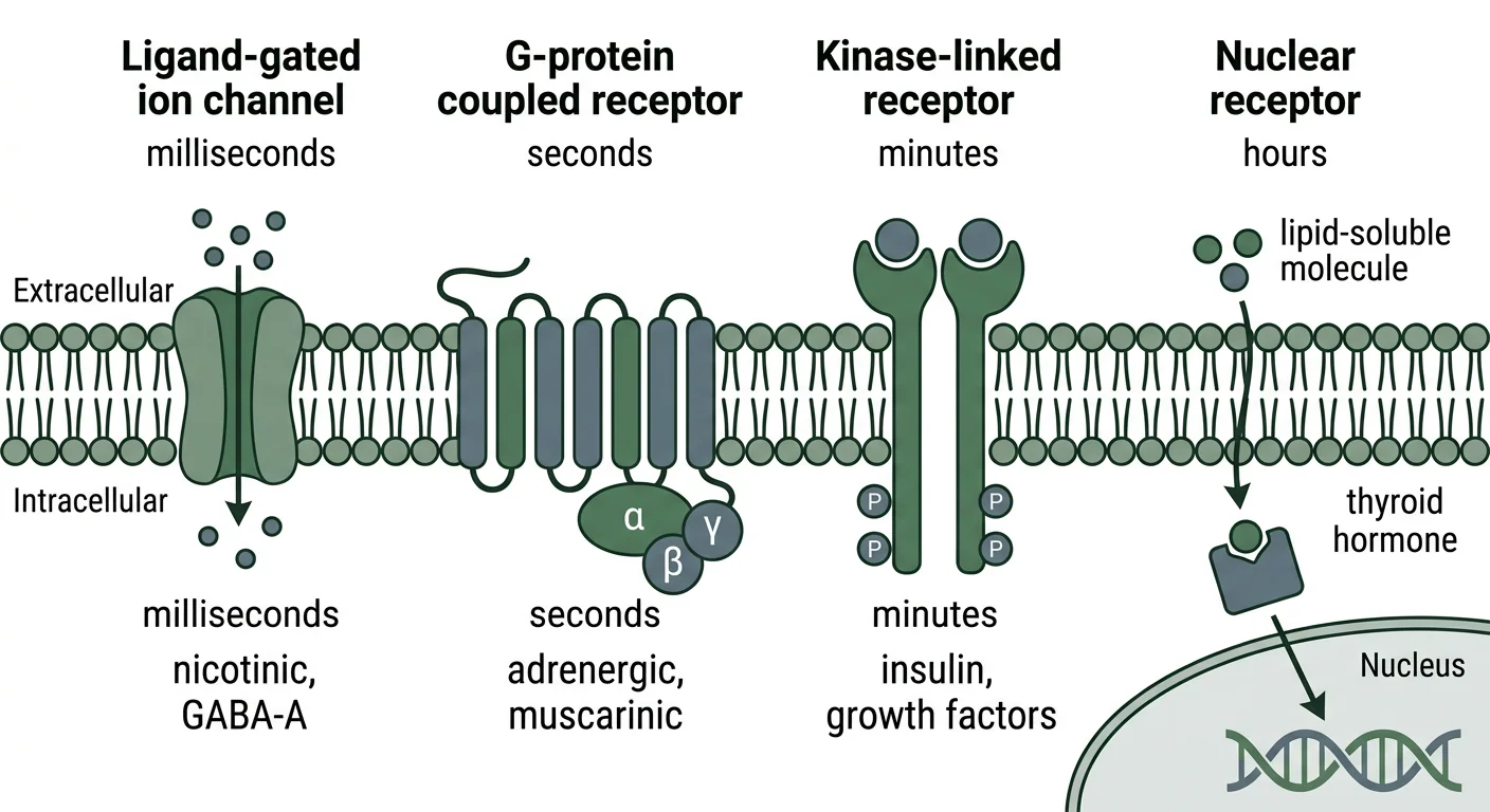
Figure 1 — The four receptor superfamilies. Diagram of the four receptor superfamilies across a cell membrane, showing ligand-gated ion channels acting in milliseconds, G-protein coupled receptors in seconds, kinase-linked receptors in minutes, and nuclear receptors acting on DNA over hours.
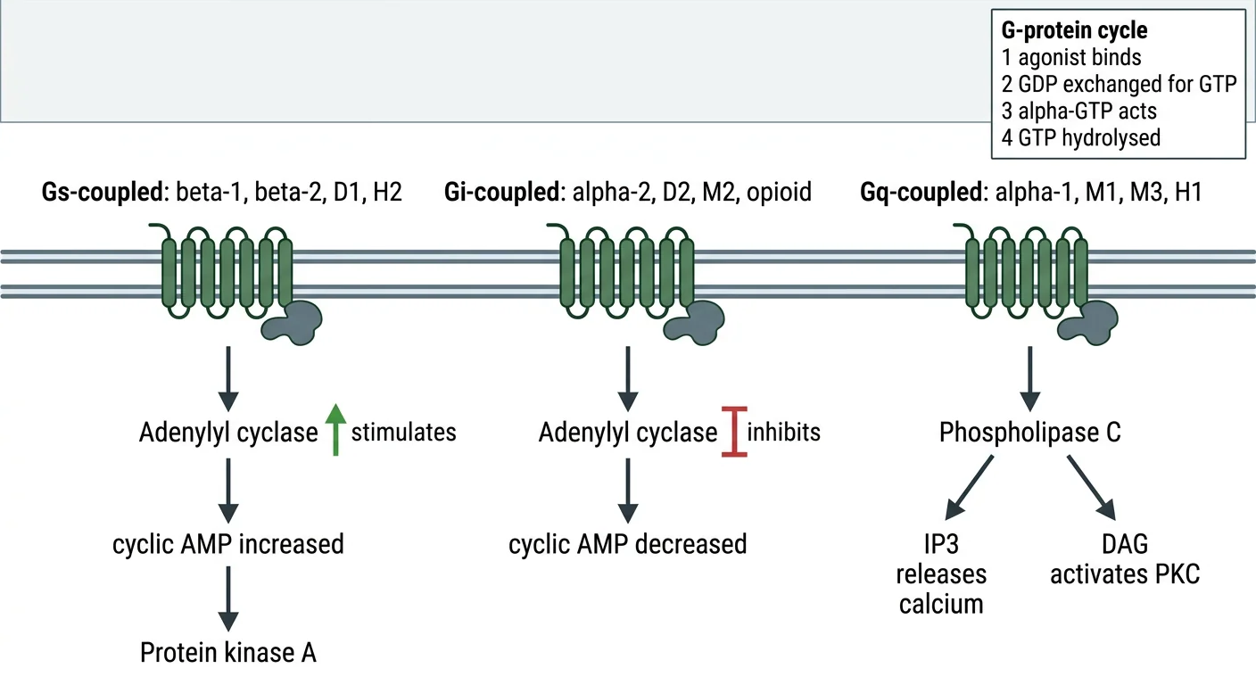
Figure 2 — G-protein signalling pathways. Diagram of the three main G-protein pathways showing Gs stimulating adenylyl cyclase to raise cyclic AMP, Gi inhibiting adenylyl cyclase to lower cyclic AMP, and Gq activating phospholipase C to generate IP3 and DAG, with an inset of the four-step G-protein activation cycle.
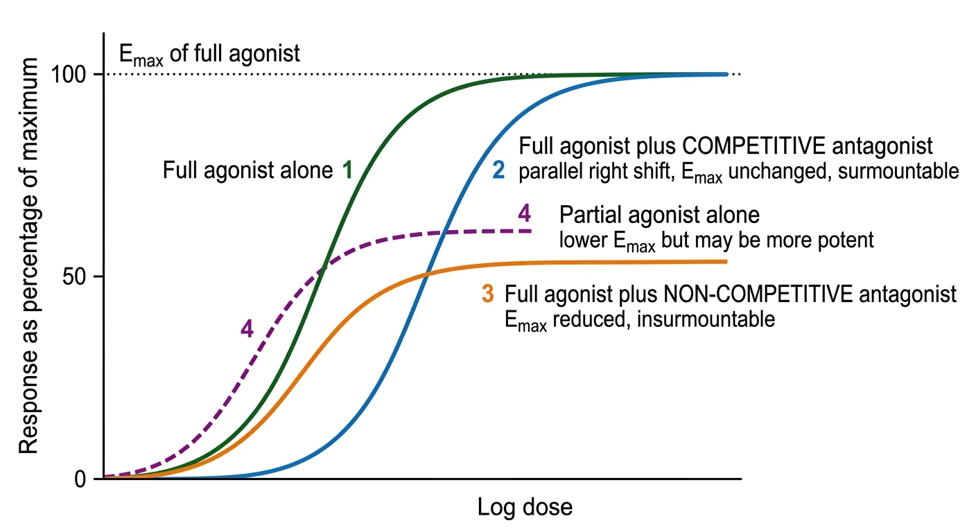
Figure 3 — Log dose–response curves: agonists and antagonists. Log dose-response graph showing a full agonist reaching maximal response, a competitive antagonist producing a parallel rightward shift with the same maximum, a non-competitive antagonist producing a lower maximum, and a partial agonist plateauing below the full agonist while lying slightly to its left.
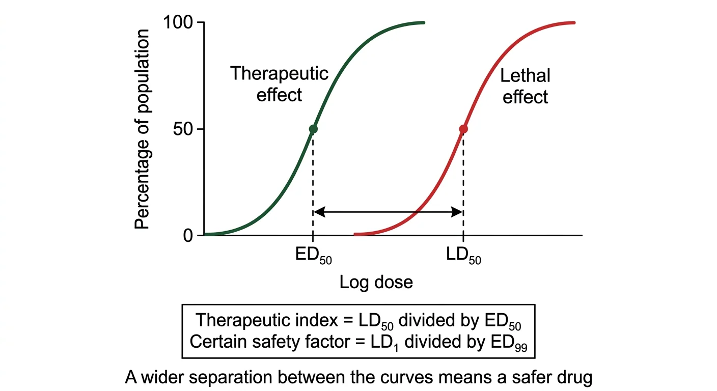
Figure 4 — Quantal dose–response curves and therapeutic index. Quantal dose-response graph showing separate cumulative curves for therapeutic and lethal effects, with ED50 and LD50 marked on the dose axis and the therapeutic index defined as their ratio.
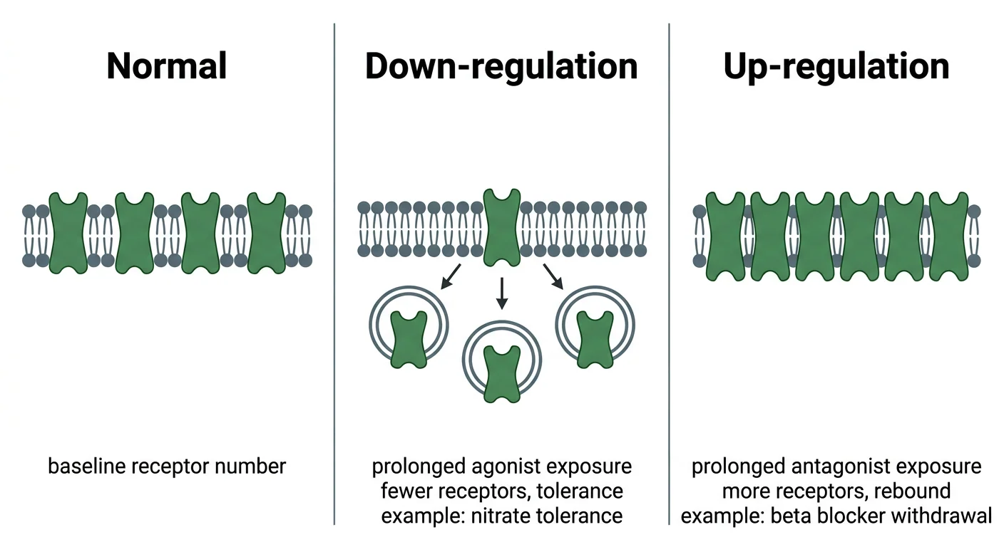
Figure 5 — Receptor regulation. Three-panel diagram contrasting normal receptor density with down-regulation from prolonged agonist exposure causing tolerance, and up-regulation from prolonged antagonist exposure causing rebound phenomena on withdrawal.
Clinical Correlation
Vignette 1 — Naloxone in opioid overdose
A 24-year-old man is brought in unconscious with pinpoint pupils and a respiratory rate of 5 per minute. Intravenous naloxone 0.4 mg produces rapid recovery of consciousness and respiration. Forty-five minutes later he becomes drowsy again with falling respiratory rate.
Reasoning: Naloxone is a competitive antagonist at the μ opioid receptor. It binds reversibly to the same site as morphine and displaces it by mass action, which is why the reversal is immediate and complete. Two properties of competitive antagonism explain the clinical course. First, the block is surmountable: a very large opioid overdose may require repeated or larger naloxone doses to shift the equilibrium sufficiently. Second, naloxone has a shorter half-life (about 60 minutes) than morphine or, especially, methadone, so as naloxone is cleared the opioid still present re-occupies the receptors and the patient re-sedates. Patients must therefore be observed, and a naloxone infusion may be required. Note also that in an opioid-dependent patient, abrupt full reversal precipitates an acute withdrawal syndrome, so titration in small increments is preferred.
Vignette 2 — Buprenorphine precipitating withdrawal
A patient maintained on high-dose morphine is given buprenorphine for pain and within an hour develops agitation, sweating, abdominal cramps, lacrimation and piloerection.
Reasoning: Buprenorphine is a partial agonist at the μ receptor with high affinity but submaximal intrinsic activity. Because its affinity exceeds that of morphine, it displaces morphine from the receptors; but because its intrinsic activity is lower, the receptors it now occupies generate a weaker signal than before. The net effect is a sudden fall in opioid receptor activation — pharmacologically identical to partial withdrawal — and the patient develops an acute abstinence syndrome. This illustrates the general rule that a partial agonist behaves as an antagonist in the presence of a full agonist. The same property confers buprenorphine's ceiling effect on respiratory depression, which makes it comparatively safer in overdose and useful in opioid substitution therapy, provided it is started only after the patient is already in mild withdrawal.
Vignette 3 — Phenoxybenzamine before surgery for phaeochromocytoma
A patient with a phaeochromocytoma is prepared for surgery with phenoxybenzamine for 10–14 days before operation, and a β-blocker is added only afterwards.
Reasoning: Phenoxybenzamine is an irreversible, non-competitive α-adrenergic antagonist that binds covalently to the receptor. Two consequences follow. Its blockade is insurmountable, so even the enormous surges of catecholamine released during tumour handling cannot overcome it — precisely the property required. And because the bond is covalent, the effect outlasts the drug's presence in plasma and ends only as new receptors are synthesised, which is why its action persists for days. The sequence matters critically: α-blockade must precede β-blockade. If a β-blocker were given first, β₂-mediated vasodilatation would be removed while α₁-mediated vasoconstriction remained unopposed, producing a hypertensive crisis. This vignette also illustrates the therapeutic value of choosing an irreversible over a competitive antagonist when insurmountable blockade is desirable.
Vignette 4 — Rebound angina after stopping a β-blocker
A patient with stable angina on long-term atenolol runs out of tablets and stops abruptly. Three days later he develops severe angina at rest and a tachycardia.
Reasoning: Prolonged receptor blockade produces adaptive up-regulation, an increase in the number of β-adrenergic receptors on the cell surface. While the antagonist is present this is silent. When it is abruptly withdrawn, the now-supranormal population of receptors is exposed to normal circulating catecholamines, producing an exaggerated response — tachycardia, hypertension, worsening angina, arrhythmia and, in susceptible patients, myocardial infarction. β-blockers must therefore always be tapered over one to two weeks rather than stopped abruptly. The identical mechanism underlies rebound hypertension after abrupt clonidine withdrawal, and the general phenomenon is the mirror image of the down-regulation that causes nitrate tolerance.
Practical Linkage
Spotter — identify the curve
| Observation | Identification | Reasoning |
|---|---|---|
| Curve reaches 100% maximum, positioned furthest left | Full agonist, most potent | Highest efficacy, lowest ED₅₀ |
| Same maximum, shifted parallel to the right | Competitive antagonist added | Emax preserved, potency reduced, surmountable |
| Maximum reduced to 50%, no rightward shift | Non-competitive antagonist added | Insurmountable, Emax reduced |
| Given alone, produces a submaximal plateau | Partial agonist | Produces a response alone, so cannot be an antagonist |
Linked competencies: PH1.5 — see also PR06 (Experimental pharmacology, computer-assisted dose–response exercises).
Exercise A — Interpreting a dose–response curve
Students are shown a graph with four labelled log dose–response curves and asked to identify each and justify the answer.
Exercise B — Calculating the therapeutic index
Drug X: ED₅₀ = 10 mg, LD₅₀ = 500 mg. Drug Y: ED₅₀ = 2 mg, LD₅₀ = 20 mg.
Solution: TI of X = 500 ÷ 10 = 50. TI of Y = 20 ÷ 2 = 10. Drug X is safer despite being less potent, and Drug Y is more potent despite being less safe. This exercise makes concrete the fact that potency and safety are independent properties.
Exercise C — Potency versus efficacy from a graph
Given three curves — drug A plateauing at 100% with ED₅₀ 8 mg, drug B plateauing at 100% with ED₅₀ 2 mg, and drug C plateauing at 60% with ED₅₀ 1 mg — rank by potency and by efficacy.
Expected answer: Potency (lowest ED₅₀ first): C > B > A. Efficacy (highest plateau first): A = B > C. Drug C is the most potent but the least efficacious, and is a partial agonist.
Exercise D — Predicting the curve
For each pair, state the expected change in the agonist curve: isoprenaline plus propranolol; acetylcholine plus atropine; noradrenaline plus phenoxybenzamine; morphine plus naloxone; GABA plus diazepam.
Expected answer: The first, second and fourth are competitive antagonists producing a parallel rightward shift with unchanged Emax. Phenoxybenzamine is irreversible and reduces Emax. Diazepam is a positive allosteric modulator and shifts the GABA curve to the left, increasing sensitivity.
MCQ Bank
40 questions · tagged by topic, exam pattern & difficulty · full explanations
Which statement about the general principles of drug action is CORRECT?
Rapid Revision
- Definition of pharmacodynamics — The study of what the drug does to the body
- Property determining whether a drug binds its receptor — Affinity, quantified by the dissociation constant Kd
- Property determining whether a bound drug activates the receptor — Intrinsic activity or efficacy
- Intrinsic activity of a full agonist, antagonist and partial agonist — One, zero and between zero and one respectively
- Drug with negative intrinsic activity — Inverse agonist
- Four receptor superfamilies in order of speed — Ligand-gated ion channels, G-protein coupled, kinase-linked, nuclear
- Timescale of ligand-gated ion channels — Milliseconds
- Timescale of nuclear receptors — Hours, because new protein must be synthesised
- Largest receptor superfamily — G-protein coupled receptors
- Effect of Gs stimulation — Adenylyl cyclase activated, cyclic AMP increased, protein kinase A activated
- Effect of Gi stimulation — Adenylyl cyclase inhibited, cyclic AMP decreased
- Effect of Gq stimulation — Phospholipase C activated, IP3 and DAG generated, calcium released
- Receptors coupled to Gq — Alpha-1, M1, M3, H1, V1 and 5-HT2
- Receptors coupled to Gi — Alpha-2, D2, M2 and the opioid receptors
- Mechanism of cholera toxin — ADP-ribosylates Gs, abolishing GTPase activity and causing persistent cyclic AMP elevation
- Mechanism of pertussis toxin — Inhibits Gi
- Second messenger releasing calcium from intracellular stores — Inositol trisphosphate
- Second messenger activating protein kinase C — Diacylglycerol
- Prototype receptor tyrosine kinase — The insulin receptor
- Pathway used by cytokine receptors — JAK-STAT
- Enzyme activated by nitric oxide — Soluble guanylyl cyclase, raising cyclic GMP
- Classic positive allosteric modulator — Benzodiazepines at the GABA-A receptor
- Why benzodiazepines are safer than barbiturates in overdose — They only modulate the response to endogenous GABA rather than opening the channel directly
- Curve change with a competitive antagonist — Parallel rightward shift with Emax unchanged, surmountable
- Curve change with a non-competitive antagonist — Emax reduced, insurmountable
- Signature of a partial agonist given alone — A submaximal plateau, but a definite response
- Why a partial agonist can precipitate opioid withdrawal — It displaces the full agonist and generates a weaker signal
- Potency on a dose-response curve — Position along the dose axis; further left means more potent
- Efficacy on a dose-response curve — Height of the plateau
- Which matters more clinically, potency or efficacy — Efficacy
- Graded curve versus quantal curve — Intensity in an individual versus proportion responding in a population
- Formula for therapeutic index — LD50 divided by ED50
- Formula for certain safety factor — LD1 divided by ED99
- Phenomenon of maximal response at partial receptor occupancy — Spare receptors or receptor reserve
- Rapidly developing tolerance over minutes to hours — Tachyphylaxis
- Consequence of prolonged agonist exposure — Down-regulation and tolerance
- Consequence of prolonged antagonist exposure — Up-regulation and rebound on withdrawal
- Reason a nitrate-free interval is prescribed — To prevent tolerance from continuous exposure
- Example of physiological antagonism — Adrenaline against histamine in anaphylaxis
- Example of chemical antagonism — Protamine neutralising heparin
- Example of synergism by sequential blockade — Sulfamethoxazole with trimethoprim
Viva Questions
- Define pharmacodynamics — The study of the biochemical and physiological effects of drugs and their mechanisms of action, that is what the drug does to the body.
- Differentiate affinity from intrinsic activity — Affinity is the tendency to bind the receptor; intrinsic activity is the ability to activate it once bound.
- Define a receptor — A specific macromolecule, usually a protein, with which a drug interacts to produce a characteristic biological response.
- Name the four receptor superfamilies with their timescales — Ligand-gated ion channels in milliseconds, G-protein coupled in seconds, kinase-linked in minutes, nuclear in hours.
- Describe the G-protein cycle — Agonist binding promotes exchange of GDP for GTP on the alpha subunit, which dissociates from beta-gamma, both regulate effectors, and intrinsic GTPase activity terminates the signal.
- Which receptors couple to Gq and what is the consequence — Alpha-1, M1, M3, H1 and 5-HT2; phospholipase C generates IP3 and DAG, raising calcium and activating protein kinase C.
- How does cholera toxin cause diarrhoea — It ADP-ribosylates Gs, abolishing GTPase activity so adenylyl cyclase is permanently active and cyclic AMP drives chloride and water secretion.
- Why do corticosteroids take hours to act — They act on nuclear receptors and must alter gene transcription and protein synthesis.
- Define a partial agonist and give an example — A drug with affinity but submaximal intrinsic activity, producing a lower maximal response than a full agonist; buprenorphine.
- Why does buprenorphine precipitate withdrawal in an opioid-dependent patient — Its high affinity displaces the full agonist while its lower intrinsic activity generates a weaker signal.
- Differentiate potency from efficacy — Potency is the dose required for a given effect, shown by the position of the curve; efficacy is the maximum achievable effect, shown by the height of the plateau.
- How does a competitive antagonist alter the dose-response curve — Parallel rightward shift, maximum unchanged, and the block is surmountable by more agonist.
- How does an irreversible antagonist differ — It reduces the maximum, is insurmountable, and its action outlasts the drug because new receptors must be synthesised.
- Define therapeutic index and state its limitation — LD50 divided by ED50; it compares only the midpoints of the two curves and ignores their slopes, which the certain safety factor addresses.
- What are spare receptors — Receptors surplus to the number needed for a maximal response, which increase tissue sensitivity to low agonist concentrations.
- Explain why a beta-blocker must be tapered — Prolonged blockade up-regulates receptors, and abrupt withdrawal exposes them to catecholamines causing rebound tachycardia and angina.
- Give one example each of physiological and chemical antagonism — Adrenaline against histamine in anaphylaxis; protamine neutralising heparin.
References
- Tripathi KD. Essentials of Medical Pharmacology. 9th ed. New Delhi: Jaypee Brothers Medical Publishers; Chapter 4 (Pharmacodynamics: Mechanism of Drug Action, Receptor Pharmacology).
- Katzung BG, Vanderah TW (eds). Basic & Clinical Pharmacology. 16th ed. New York: McGraw Hill; Chapters 1 and 2 (Introduction; Drug Receptors and Pharmacodynamics).
- Brunton LL, Knollmann BC (eds). Goodman & Gilman's The Pharmacological Basis of Therapeutics. 14th ed. New York: McGraw Hill; Chapter 3 (Pharmacodynamics: Molecular Mechanisms of Drug Action).
- Ritter JM, Flower RJ, Henderson G, et al. Rang & Dale's Pharmacology. 10th ed. Edinburgh: Elsevier; Chapters 2 and 3 (How Drugs Act: General Principles; Molecular Aspects).
- National Medical Commission. Competency Based Undergraduate Curriculum for the Indian Medical Graduate, Volume II — Pharmacology (Code: PH), competency PH1.5.
- Alexander SPH, Christopoulos A, Davenport AP, et al. The Concise Guide to Pharmacology: G protein-coupled receptors. British Journal of Pharmacology (IUPHAR/BPS Guide to Pharmacology), current edition.
- Kenakin T. A Pharmacology Primer: Techniques for More Effective and Strategic Drug Discovery. 5th ed. London: Academic Press; chapters on receptor theory, allosterism and inverse agonism.
- Sharma HL, Sharma KK. Principles of Pharmacology. 4th ed. Hyderabad: Paras Medical Publisher; chapters on receptors and dose-response relationships.
Downloads
All deliverables & printable revision sheets for CH04
Printable Chapter Handout & Study Notes
Generate a clean, high-yield PDF or print directly without sidebars and navigation menus.