
Pharmacokinetics I — Absorption, Bioavailability, Distribution
Learning Objectives
At the end of this chapter, the Phase II MBBS student will be able to:
- Enumerate and describe the mechanisms by which drugs cross biological membranes, and distinguish passive diffusion from carrier-mediated transport. (PH1.4 — Knows)
- State the pH partition hypothesis and apply the Henderson–Hasselbalch equation to predict the ionisation of a weak acid or weak base at a given pH. (PH1.4 — Knows-how)
- Explain ion trapping and apply it to the gastric absorption of drugs, to urinary alkalinisation in poisoning, and to drug transfer into breast milk. (PH1.4 — Knows-how)
- Analyse the physicochemical, pharmaceutical and physiological factors that modify drug absorption. (PH1.4 — Knows-how)
- Define bioavailability and distinguish it from bioequivalence and therapeutic equivalence, and calculate absolute bioavailability from area-under-the-curve data. (PH1.4 — Shows-how)
- Describe the determinants of drug distribution, including perfusion, lipid solubility and tissue binding. (PH1.4 — Knows)
- Define apparent volume of distribution, calculate it from dose and plasma concentration, and interpret high and low values in clinical terms. (PH1.4 — Shows-how)
- Explain the consequences of plasma protein binding and predict when a displacement interaction will be clinically significant. (PH1.4 — Knows-how)
- Describe the blood–brain and placental barriers, the role of P-glycoprotein, and the phenomenon of redistribution. (PH1.4 — Knows)
- Solve numerical problems on volume of distribution, loading dose, percentage ionisation and bioavailability. (PH1.4, PH2.4 — Shows-how)
Must-Know Summary
Pharmacokinetics is what the body does to the drug. It has four processes — absorption, distribution, metabolism and excretion — and every one of them requires the drug to cross a lipid membrane. This chapter deals with the first half of that story: how drugs get in, and where they go once they are in.
Two ideas carry most of the clinical weight. The first is that only the unionised, lipid-soluble form of a drug crosses membranes, so the pH of a compartment relative to the drug's pKa determines both where it is absorbed and where it accumulates. The second is that the volume of distribution is a calculated proportion, not a real anatomical space — it tells you how much of the drug is hiding outside the plasma, and therefore whether a loading dose is needed and whether dialysis will work.
In one line each:
- Most drugs cross membranes by passive diffusion of the unionised form, down a concentration gradient, without energy or a carrier.
- Carrier-mediated transport is saturable, structurally specific and subject to competitive inhibition; passive diffusion is none of these.
- Only the unionised form is lipid-soluble and diffusible; the ionised form is trapped.
- Weak acids are relatively unionised in the acidic stomach and can be absorbed there; weak bases are unionised in the alkaline intestine.
- When pH equals pKa, the drug is exactly 50% ionised.
- Alkalinising the urine accelerates excretion of a weak acid such as salicylate or phenobarbitone — the ionised drug cannot be reabsorbed.
- Bioavailability is the fraction reaching the systemic circulation unchanged; F = AUC(oral)/AUC(IV).
- Vd = Dose / plasma concentration. It is apparent — it may vastly exceed total body water.
- A high Vd means the drug is sequestered in tissue, so it is not removed by dialysis; a low Vd with low protein binding is dialysable.
- Warfarin is highly lipid-soluble but has a low Vd, because 99% plasma protein binding holds it in the vascular compartment — binding beats lipophilicity.
- Acidic drugs bind albumin; basic drugs bind α1-acid glycoprotein.
- Sulfonamides displace bilirubin from albumin in the neonate and can cause kernicterus.
- The brief action of thiopentone is terminated by redistribution from brain to muscle and fat, not by metabolism.
Classification
Box 1 — Mechanisms of drug transport across biological membranes
I. Passive (no energy, no carrier, down the gradient)
- Passive (simple) diffusion of the unionised lipid-soluble form — the major mechanism for most drugs
- Filtration / bulk flow through aqueous pores and fenestrations — small water-soluble molecules
- Diffusion through ion channels — a few small ions
II. Carrier-mediated (saturable, specific, competitively inhibited)
- Facilitated diffusion — carrier used, gradient followed, no energy required
- Active transport — against the gradient, energy required
- Primary active transport: ATP hydrolysed directly (Na⁺/K⁺-ATPase; ABC efflux pumps)
- Secondary active transport: coupled to an ion gradient — symport or antiport
- Efflux transporters: P-glycoprotein (MDR1), BCRP, MRP family
III. Vesicular transport
- Pinocytosis and receptor-mediated endocytosis — large molecules, proteins, monoclonal antibodies
Box 2 — Determinants of drug distribution
Drug factors
- Lipid solubility and partition coefficient
- Degree of ionisation at physiological pH (pKa)
- Molecular size
- Affinity for plasma proteins
- Affinity for tissue constituents
Body factors
- Regional blood flow and perfusion
- Presence of specialised barriers (blood–brain, blood–CSF, placental, blood–testis)
- Body composition — water, fat and muscle mass; age, sex, obesity, pregnancy
- Plasma protein concentration — albumin and α1-acid glycoprotein
- Disease states — oedema, cirrhosis, nephrotic syndrome, inflammation
Core Concepts
1. Biological membranes and mechanisms of drug transport
Every pharmacokinetic process — absorption from the gut, distribution into tissue, reabsorption in the renal tubule, entry into a hepatocyte for metabolism — requires the drug molecule to traverse at least one biological membrane. The membrane is a phospholipid bilayer roughly 10 nm thick with its hydrophobic hydrocarbon core sandwiched between hydrophilic polar surfaces, interrupted by integral proteins that serve as channels, carriers and pumps.
The consequence is simple and governs everything that follows: a lipid-soluble, uncharged, small molecule crosses easily; a water-soluble, charged, large molecule does not.
Passive diffusion is quantitatively the most important mechanism for the great majority of drugs. The drug moves down its concentration gradient, without expenditure of energy and without a carrier, at a rate described by Fick's law — proportional to the concentration gradient, the surface area available and the lipid–water partition coefficient, and inversely proportional to membrane thickness. Because there is no carrier, passive diffusion is not saturable, not structurally specific and not subject to competitive inhibition. Only the unionised fraction diffuses, which is why the pH partition principle discussed below is so important.
Filtration (bulk flow through aqueous channels) allows very small water-soluble molecules (molecular weight under about 100–200 Da, such as urea, ethanol and water itself) to pass through aqueous pores in the membrane, and permits considerably larger molecules to cross the fenestrated capillary endothelium of most tissues. Glomerular filtration is the clearest example.
Carrier-mediated transport uses a specific transmembrane protein. All carrier systems share three defining features, which distinguish them from passive diffusion in examination questions:
- Saturability — a finite number of carriers gives a maximum transport rate (Tmax).
- Structural specificity — the carrier recognises a particular molecular shape.
- Competitive inhibition — structurally similar substrates compete for the same carrier.
Two subtypes exist. In facilitated diffusion, transport follows the concentration gradient and requires no energy; the carrier merely speeds a thermodynamically favourable movement (e.g. glucose entry via GLUT transporters, vitamin B12 absorption with intrinsic factor). In active transport, the drug moves against its electrochemical gradient and energy is therefore required. Primary active transport hydrolyses ATP directly (the Na⁺/K⁺-ATPase, and the ABC family of efflux pumps). Secondary active transport couples the movement of the drug to the downhill movement of an ion, either in the same direction (symport, e.g. the sodium-dependent glucose transporter) or in the opposite direction (antiport, e.g. the Na⁺/H⁺ exchanger). Levodopa, methyldopa, 5-fluorouracil and many beta-lactams are handled by amino acid or peptide transporters.
Efflux transporters deserve separate emphasis because they are increasingly examined. P-glycoprotein (P-gp, the product of the ABCB1/MDR1 gene) is an ATP-dependent pump expressed on the apical membrane of intestinal enterocytes, the biliary canaliculus, the renal proximal tubule, the placenta and — critically — the luminal surface of brain capillary endothelium. It extrudes a structurally diverse range of substrates back into the gut lumen, bile, urine or blood, thereby reducing oral bioavailability and restricting entry into the brain and fetus. Digoxin, ciclosporin, many anticancer drugs, loperamide and several protease inhibitors are substrates; verapamil, quinidine, ciclosporin, ketoconazole and clarithromycin inhibit it, while rifampicin induces it. Over-expression of P-gp in tumour cells is one mechanism of multidrug resistance. Related pumps include BCRP (breast cancer resistance protein) and the MRP family, while the OATP uptake transporters move drugs such as statins into hepatocytes.
Pinocytosis and endocytosis account for the cellular uptake of a small number of very large molecules and particles — proteins, monoclonal antibodies, vitamin B12 complexes, and iron bound to transferrin.
Ionic or electrochemical channels permit passage of a few very small ions (lithium, for instance, enters through sodium channels).
2. The pH partition hypothesis and ionisation
Most drugs are either weak acids or weak bases and therefore exist in solution as an equilibrium between an unionised and an ionised species. The pH partition hypothesis states that only the unionised form, being lipid-soluble, can cross a biological membrane by passive diffusion; the ionised form, being charged and hydrophilic, cannot.
The proportion of each species is given by the Henderson–Hasselbalch equation.
For a weak acid (HA ⇌ H⁺ + A⁻, where HA is unionised):
pH = pKa + log ([ionised] / [unionised]) = pKa + log ([A⁻] / [HA])
For a weak base (BH⁺ ⇌ B + H⁺, where B is unionised):
pH = pKa + log ([unionised] / [ionised]) = pKa + log ([B] / [BH⁺])
Three consequences follow, and they are the basis of most examination questions on this topic:
- When pH = pKa, the drug is exactly 50% ionised. This is the definition of pKa.
- When the pH differs from the pKa by one unit, the ratio is 10:1 — that is, approximately 91% in one form and 9% in the other. A difference of two units gives roughly 99:1.
- A weak acid is progressively more ionised as the medium becomes more alkaline; a weak base is more ionised as the medium becomes more acidic. The mnemonic that helps is that like dissolves like but like does not ionise in like: an acid placed in an acidic medium stays unionised and is therefore absorbable.
Application to absorption. Aspirin is a weak acid with a pKa of about 3.5. In gastric juice at pH 2 it is largely unionised and therefore lipid-soluble, and can be absorbed across the gastric mucosa. Morphine and atropine are weak bases and are almost completely ionised at gastric pH, so they are not absorbed from the stomach; they are absorbed in the small intestine where the pH is higher.
It is important, however, not to over-apply the principle. In practice the small intestine is the major site of absorption for almost all orally administered drugs — including weak acids — because its enormous surface area (villi and microvilli) and rich blood supply overwhelm the theoretical pH advantage of the stomach. The pH partition principle predicts the direction of a pH effect; surface area determines the magnitude of actual absorption.
Ion trapping is the accumulation of a drug on one side of a membrane because the pH there converts it to the ionised, non-diffusible form. The unionised molecule diffuses across, becomes ionised, and cannot diffuse back. Clinically important examples:
- Gastric mucosal injury by aspirin. Unionised aspirin diffuses into the gastric mucosal cell, where the intracellular pH of about 7.0 ionises it. The trapped anion accumulates and contributes to local mucosal damage, in addition to the systemic COX-1 effect.
- Urinary trapping in poisoning (discussed below).
- Breast milk. Milk is slightly more acidic (pH about 6.8–7.0) than plasma (7.4), so weak bases are concentrated in milk relative to plasma, while weak acids are relatively excluded.
- The fetus. Fetal blood is slightly more acidic than maternal blood, so weak bases such as local anaesthetics can be trapped in the fetal circulation — a genuine concern with prolonged epidural anaesthesia and one reason for fetal acidosis worsening local anaesthetic accumulation.
- Abscess cavities. Inflamed and infected tissue is acidic, which ionises local anaesthetics (weak bases) and reduces the unionised fraction available to cross the nerve membrane. This is the principal reason local anaesthesia often fails in infected tissue.
Therapeutic exploitation in poisoning. Because only the unionised drug is passively reabsorbed from the renal tubular fluid, manipulating urinary pH alters excretion:
- Alkalinising the urine with intravenous sodium bicarbonate to a target urine pH of about 7.5–8.0 traps weak acids in the tubular lumen and enhances their elimination. This is established therapy in salicylate poisoning and is also used for phenobarbitone and methotrexate toxicity.
- Acidifying the urine would, by the same logic, enhance elimination of weak bases such as amphetamine, phencyclidine and quinine. This is a valid pharmacological principle but is no longer recommended in clinical practice, because acidification risks worsening metabolic acidosis and precipitating myoglobinuric renal failure in patients who are frequently already rhabdomyolytic. Students should know the principle and its examples, and should also know that it is not current standard care.
A note on nuance. The classical teaching is that urinary alkalinisation works by increasing ionisation of salicylate. Some authors point out that salicylic acid (pKa ≈ 3.0–3.5) is already almost entirely ionised at any physiological urinary pH, so further alkalinisation cannot increase ionisation much; alternative explanations invoke partitioning of the unionised fraction from peritubular fluid into the lumen. The clinically and examination-relevant answer remains ion trapping, but the mechanism is not as arithmetically tidy as the standard diagram suggests.
3. Absorption and the factors that modify it
Absorption is the movement of a drug from its site of administration into the systemic circulation. It is complete and instantaneous only after intravenous injection; by every other route it is a variable that determines both the rate of onset and the amount that ultimately arrives.
Physicochemical factors of the drug
- Lipid solubility — the single most important determinant; highly lipid-soluble drugs are well absorbed.
- Degree of ionisation (pKa) and the pH at the absorption site — as discussed above.
- Molecular size — very large molecules are poorly absorbed orally; peptides are additionally destroyed by proteases.
- Aqueous solubility and dissolution rate — a drug must dissolve before it can be absorbed. Very insoluble drugs and very rapidly precipitating ones (phenytoin injected intramuscularly) are absorbed erratically.
- Particle size — smaller particles dissolve faster; micronised griseofulvin and aspirin are absorbed better than coarse preparations.
Pharmaceutical factors — disintegration and dissolution time of the tablet, the nature of the excipients, the salt form used, and the presence of a coating. Two products containing the same amount of the same drug may be absorbed very differently, which is exactly why bioequivalence testing exists.
Physiological and patient factors
- Surface area and vascularity of the absorbing surface — the small intestine dominates for this reason.
- Splanchnic blood flow — reduced in shock and after a meal in some vascular beds; absorption falls when perfusion falls.
- Gastric emptying rate — because absorption mainly occurs distal to the stomach, anything that accelerates gastric emptying accelerates absorption (metoclopramide) and anything that delays it slows absorption (opioids, anticholinergics, food, migraine itself).
- Gastrointestinal motility — very rapid transit (diarrhoea, purgatives) reduces the contact time available for absorption.
- Presence of food — usually delays and may reduce absorption (most antibiotics, and notably levothyroxine and bisphosphonates, which must be taken on an empty stomach); occasionally food increases absorption of highly lipophilic drugs (griseofulvin, albendazole, saquinavir) by stimulating bile secretion.
- Chelation and adsorption interactions — tetracyclines and fluoroquinolones chelate calcium, magnesium, aluminium and iron, so milk and antacids markedly reduce their absorption. Cholestyramine adsorbs digoxin, warfarin and thyroxine.
- Gut wall metabolism and efflux — CYP3A4 in the enterocyte and P-glycoprotein both reduce the fraction reaching the portal blood. Grapefruit juice inhibits intestinal CYP3A4 and thereby raises the bioavailability of felodipine and several other drugs.
- Gut flora — bacteria in the colon deconjugate drugs and participate in enterohepatic recycling; antibiotics that suppress flora can reduce the efficacy of oral contraceptives by this route.
- Disease states — malabsorption syndromes, coeliac disease, achlorhydria, previous gastric surgery and heart failure with gut oedema all alter absorption.
4. Bioavailability, bioequivalence and therapeutic equivalence
Bioavailability (F) is the fraction of an administered dose that reaches the systemic circulation in the unchanged, active form. By definition F = 1 (100%) for an intravenous dose. For all other routes F is reduced by incomplete absorption and by pre-systemic (first-pass) elimination.
Measurement. The area under the plasma concentration–time curve (AUC) is proportional to the total amount of drug reaching the circulation. Hence:
Absolute bioavailability, F = AUC(oral) / AUC(IV) — for the same dose. If different doses are used, each AUC is corrected for dose.
Relative bioavailability compares a test formulation with a reference non-intravenous formulation: F(rel) = AUC(test) / AUC(reference).
Two further parameters describe the rate rather than the extent of absorption: Cmax, the peak plasma concentration, and Tmax, the time at which it occurs. A formulation may deliver the same total amount of drug (same AUC) more slowly (lower Cmax, later Tmax) — which is precisely what a sustained-release product is designed to do.
Bioequivalence means that two formulations of the same drug, given in the same dose, produce plasma concentration–time profiles so similar that their therapeutic effects can be expected to be essentially the same. Regulatory bioequivalence conventionally requires the 90% confidence interval of the ratio of AUC and Cmax to fall within 80–125% of the reference product.
Therapeutic equivalence is the broader clinical claim: the two products, given to the same patient, produce the same therapeutic effect and the same safety profile. Bioequivalence is normally accepted as a surrogate for it.
When does this matter clinically? For most drugs a modest difference in bioavailability is inconsequential. It becomes critical for drugs with a narrow therapeutic index, where a small change in plasma concentration moves the patient between subtherapeutic and toxic — warfarin, phenytoin, digoxin, lithium, levothyroxine, ciclosporin, tacrolimus and carbamazepine. For these, switching brands should be avoided or accompanied by monitoring.
5. Distribution: compartments and determinants
Distribution is the reversible movement of drug from the systemic circulation into the tissues. Its extent is determined by four principal factors.
- Regional blood flow. Well-perfused organs — brain, heart, liver, kidney, endocrine glands — receive drug within minutes; poorly perfused tissues such as fat, resting skeletal muscle, bone and skin equilibrate over hours. This perfusion difference is the basis of redistribution.
- Lipid solubility and molecular characteristics. Lipophilic unionised drugs cross into cells and accumulate in fat; hydrophilic or highly ionised drugs are largely confined to the extracellular fluid. Aminoglycosides and neuromuscular blockers, being highly polar, are essentially restricted to the extracellular compartment.
- Plasma protein binding. Bound drug cannot leave the vascular compartment; extensive binding restricts distribution (see below).
- Tissue binding and sequestration. Drugs may concentrate in specific tissues far above plasma levels: digoxin in cardiac and skeletal muscle (Na⁺/K⁺-ATPase), chloroquine in liver, spleen and retina (melanin binding, hence retinopathy), tetracyclines and heavy metals in bone and teeth, iodine in thyroid, amiodarone in fat, lung and cornea, and griseofulvin in keratin.
For reference, the body fluid compartments of a 70 kg adult are approximately: plasma 3 L, blood 5 L, extracellular fluid 14 L (about 0.2 L/kg), intracellular fluid 28 L, and total body water 42 L (about 0.6 L/kg), with adipose tissue contributing a further variable volume.
6. Apparent volume of distribution
The apparent volume of distribution (Vd) is the hypothetical volume of body fluid that would be required to contain the total amount of drug in the body at the same concentration as that measured in plasma.
Vd = Total amount of drug in the body / Plasma concentration
After an intravenous bolus: Vd = Dose / C₀, where C₀ is the plasma concentration extrapolated back to time zero.
It may be expressed in litres, or normalised to body weight in L/kg.
The word apparent is essential. Vd is a proportionality constant, not an anatomical space. It can be far larger than the entire body — chloroquine has a Vd of the order of 13,000 L — because the calculation simply asks "how much fluid would be needed to explain this low plasma concentration, if the drug were evenly distributed?" A very low plasma concentration for a given dose implies most of the drug is elsewhere, and the arithmetic returns a very large number.
The warfarin paradox is a favourite examination point. Warfarin is a lipid-soluble molecule, yet its Vd is small (about 8–10 L). The reason is that it is 99% bound to plasma albumin, and bound drug cannot leave the vascular compartment. Where the two properties conflict, plasma protein binding predominates over lipid solubility in determining Vd.
Clinical applications
- Loading dose. To fill the volume of distribution rapidly: Loading dose = (Target plasma concentration × Vd) / F. Note that the loading dose depends on Vd, whereas the maintenance dose depends on clearance — a distinction developed further in CH03.
- Predicting dialysability. Haemodialysis removes drug from plasma. A drug is well removed if it has a low Vd, low plasma protein binding, low molecular weight and good water solubility (lithium, methanol, ethylene glycol, salicylate, phenobarbitone, theophylline). A drug with a high Vd is poorly removed no matter how efficient the dialyser, because the overwhelming majority of the drug is in tissue and not in the plasma being filtered — digoxin, amiodarone, tricyclic antidepressants and benzodiazepines are the standard examples.
- Estimating the body burden in overdose from a measured plasma concentration.
Factors altering Vd include age (neonates have proportionately more body water; the elderly have more fat and less water), sex, obesity, pregnancy, oedematous states such as heart failure, cirrhosis and nephrotic syndrome, and dehydration.
7. Plasma protein binding
Many drugs circulate partly bound to plasma proteins. The binding is reversible and follows the law of mass action, so bound and free drug are in dynamic equilibrium.
- Acidic drugs bind principally to albumin — warfarin, phenytoin, valproate, NSAIDs, sulfonamides, penicillins, furosemide.
- Basic drugs bind principally to α1-acid glycoprotein (AAG) — lignocaine, bupivacaine, propranolol, quinidine, imipramine, disopyramide.
- Corticosteroid-binding globulin, sex hormone-binding globulin and transferrin bind specific endogenous ligands and their drug analogues.
Consequences of binding
- Only free drug is pharmacologically active. Bound drug is a temporarily inert reservoir.
- Only free drug is available for distribution, so high binding restricts Vd.
- Only free drug undergoes glomerular filtration; bound drug is not filtered (though it may still be actively secreted, as penicillin is).
- Binding prolongs duration of action by acting as a slowly releasing depot.
- Binding is saturable, so at high doses the free fraction can rise disproportionately.
Displacement interactions. One drug may displace another from its binding site, transiently increasing the free concentration of the displaced drug. The clinical importance of this mechanism has historically been overstated: for most drugs, the displaced free drug is immediately available for redistribution and elimination, so the free concentration returns towards baseline and no harm results.
A displacement interaction is clinically significant only when several conditions coincide: the displaced drug is highly bound (>90%), has a narrow therapeutic index, has a small volume of distribution, and its elimination is simultaneously impaired or slow. This is why the genuine examples are few but important:
- Sulfonamides (including cotrimoxazole) displace bilirubin from albumin in the neonate. Free bilirubin crosses the immature blood–brain barrier and is deposited in the basal ganglia, producing kernicterus. Sulfonamides are therefore contraindicated in neonates under about two months and in the third trimester of pregnancy. This is the single most examined displacement interaction in the subject.
- Aspirin and other NSAIDs, sulfonamides and valproate displace warfarin, increasing the risk of bleeding — though with warfarin, concurrent inhibition of metabolism and of platelet function usually contributes more than displacement alone.
- Valproate displaces phenytoin, raising the free phenytoin fraction. Because laboratories usually report total phenytoin, the reported level may look normal or even low while the patient is clinically toxic — a well-recognised trap. Free phenytoin measurement is preferred in this situation and in hypoalbuminaemia.
Hypoalbuminaemia — in nephrotic syndrome, cirrhosis, malnutrition, burns and old age — raises the free fraction of acidic drugs and can produce toxicity at conventional total-drug concentrations. Conversely AAG rises as an acute-phase reactant in inflammation, myocardial infarction, surgery, trauma and cancer, increasing the bound fraction of basic drugs and potentially reducing their free active concentration.
8. Special barriers, redistribution and tissue reservoirs
The blood–brain barrier (BBB) is formed by capillary endothelial cells joined by continuous tight junctions without fenestrations, invested by pericytes and the foot processes of astrocytes. It behaves as a continuous lipid barrier, so only lipid-soluble unionised drugs cross by passive diffusion; polar and ionised drugs are excluded. Specific carriers transport essential nutrients and their analogues — the large neutral amino acid carrier admits levodopa, which is why the prodrug works where dopamine cannot. P-glycoprotein on the luminal endothelial membrane actively pumps many drugs back into the blood, further restricting entry; loperamide, an opioid, produces no central effect for precisely this reason.
Two qualifications are clinically important. Certain circumventricular organs lack a barrier — notably the chemoreceptor trigger zone in the area postrema (which is why dopamine antagonists that do not otherwise enter the brain can act as antiemetics), the median eminence and the posterior pituitary. And inflammation increases permeability: in bacterial meningitis, penicillin and third-generation cephalosporins penetrate far better than they do into a healthy meninx, which is what makes therapy possible.
The blood–CSF barrier at the choroid plexus is analogous. It also possesses an active organic acid transport system that pumps drugs such as penicillin out of the CSF, an efflux that probenecid can inhibit.
The placenta does not exclude drugs effectively. It is a lipid barrier with a large surface area and rich blood flow, and lipid-soluble unionised drugs cross readily; the working assumption in clinical practice must be that any drug given to the mother reaches the fetus to some degree. Placental P-glycoprotein affords partial protection against some substrates. Heparin (large and highly polar) and insulin (a peptide) are the classic drugs that do not cross meaningfully — which is why heparin, not warfarin, is the anticoagulant of choice in pregnancy.
Redistribution is the phenomenon whereby a highly lipid-soluble drug given intravenously first enters the well-perfused brain, producing a rapid effect, and is then carried away as the drug equilibrates with less well-perfused muscle and fat, terminating the effect.
Thiopentone is the classic example. After a single intravenous induction dose it reaches the brain within one arm–brain circulation time and produces anaesthesia in seconds. Consciousness returns within 5–10 minutes — not because the drug has been metabolised or excreted, but because it has redistributed out of the brain into muscle and then fat. Its elimination half-life is many hours, and the drug is still present in the body long after the patient has woken. Two consequences follow: repeated or large doses saturate the peripheral tissues, so redistribution can no longer terminate the effect and recovery becomes prolonged (cumulation), and the effect is exaggerated and prolonged in patients with a small muscle mass. The same principle governs the short action of fentanyl and propofol boluses.
Tissue reservoirs prolong the presence of a drug in the body and can be a source of delayed toxicity: fat (thiopentone, amiodarone, organochlorine insecticides), bone and teeth (tetracyclines, lead, fluoride, bisphosphonates), the retina (chloroquine), keratin of skin, hair and nails (griseofulvin, arsenic), and cells generally (digoxin in muscle).
Tables
Table 1 — Mechanisms of transport across biological membranes
| Feature | Passive diffusion | Filtration | Facilitated diffusion | Active transport |
|---|---|---|---|---|
| Carrier required | No | No | Yes | Yes |
| Energy required | No | No | No | Yes |
| Direction | Down gradient | Down gradient | Down gradient | Against gradient |
| Saturable | No | No | Yes | Yes |
| Structural specificity | No | No | Yes | Yes |
| Competitive inhibition | No | No | Yes | Yes |
| Example | Most drugs (unionised form) | Urea, water, small ions | Glucose via GLUT; vitamin B12 | Levodopa, methyldopa, 5-FU; P-gp efflux |
Table 2 — pKa of representative drugs and preferential site of absorption
| Drug | Type | Approximate pKa | Behaviour |
|---|---|---|---|
| Aspirin | Weak acid | 3.5 | Largely unionised in stomach; can be absorbed gastrically |
| Warfarin | Weak acid | 5.0 | Highly bound to albumin; absorbed in intestine |
| Phenobarbitone | Weak acid | 7.4 | Excretion enhanced by urinary alkalinisation |
| Phenytoin | Weak acid | 8.4 | Poorly water-soluble; erratic IM absorption |
| Diazepam | Very weak base | 3.0 | Largely unionised at intestinal pH |
| Lignocaine | Weak base | 7.9 | Ionised in acidic tissue; fails in abscess |
| Morphine | Weak base | 7.9 | Ionised in stomach; absorbed in intestine |
| Amphetamine | Weak base | 9.9 | Excretion theoretically enhanced by acid urine |
Table 3 — Effect of the difference between pH and pKa on ionisation
| pH − pKa | Weak acid: % ionised | Weak acid: % unionised |
|---|---|---|
| −2 | ~1% | ~99% |
| −1 | ~9% | ~91% |
| 0 | 50% | 50% |
| +1 | ~91% | ~9% |
| +2 | ~99% | ~1% |
Table 4 — Ion trapping in clinical practice
| Situation | Drug type | Where trapped | Clinical consequence or use |
|---|---|---|---|
| Gastric mucosal cell | Weak acid (aspirin) | Inside the cell (pH 7.0) | Contributes to local mucosal injury |
| Alkaline urine | Weak acid (salicylate, phenobarbitone) | Tubular lumen | Enhanced excretion — used in poisoning |
| Acidic urine | Weak base (amphetamine, quinine) | Tubular lumen | Enhanced excretion — principle valid, not current practice |
| Breast milk (pH ~6.8) | Weak base | Milk | Basic drugs concentrate in milk |
| Fetal circulation | Weak base (local anaesthetics) | Fetus | Accumulation, worse with fetal acidosis |
| Abscess / inflamed tissue | Weak base (lignocaine) | Extracellular acidic fluid | Local anaesthetic failure |
Table 5 — Factors affecting drug absorption
| Category | Factor | Effect |
|---|---|---|
| Drug | Lipid solubility | Higher solubility, better absorption |
| Drug | Ionisation (pKa vs pH) | Only unionised form absorbed |
| Drug | Particle size, dissolution rate | Smaller particles absorbed faster |
| Formulation | Coating, excipients, salt form | Determines disintegration and dissolution |
| GI tract | Surface area | Small intestine dominant for nearly all drugs |
| GI tract | Gastric emptying | Faster emptying speeds absorption |
| GI tract | Motility | Rapid transit reduces absorption |
| GI tract | Food | Usually delays; increases absorption of lipophilic drugs |
| Interaction | Chelation | Antacids, milk, iron reduce tetracycline and quinolone absorption |
| Barrier | Gut CYP3A4 and P-glycoprotein | Reduce fraction reaching portal blood |
| Patient | Splanchnic blood flow | Reduced in shock, absorption unreliable |
| Patient | Disease | Malabsorption, achlorhydria, gut oedema alter absorption |
Table 6 — Bioavailability, bioequivalence and therapeutic equivalence
| Term | Definition | How assessed |
|---|---|---|
| Bioavailability (F) | Fraction of dose reaching systemic circulation unchanged | F = AUC(oral) / AUC(IV), same dose |
| Relative bioavailability | Comparison of a test formulation with a non-IV reference | AUC(test) / AUC(reference) |
| Bioequivalence | Two formulations give sufficiently similar concentration–time profiles | 90% CI of AUC and Cmax ratio within 80–125% |
| Therapeutic equivalence | Same clinical efficacy and safety in the patient | Bioequivalence accepted as surrogate |
Table 7 — Volume of distribution: landmark values
| Approximate Vd (70 kg adult) | Interpretation | Representative drugs |
|---|---|---|
| 3–5 L | Confined to plasma | Heparin, warfarin |
| ~14 L (0.2 L/kg) | Extracellular fluid | Gentamicin, mannitol, neuromuscular blockers |
| ~42 L (0.6 L/kg) | Total body water | Ethanol, theophylline, phenytoin |
| 100–500 L | Moderate tissue binding | Propranolol, morphine |
| > 500 L | Extensive sequestration; not dialysable | Digoxin (~500 L), amiodarone, chloroquine (very high), imipramine |
Table 8 — Plasma protein binding
| Feature | Albumin | α1-Acid glycoprotein |
|---|---|---|
| Binds | Acidic and neutral drugs | Basic drugs |
| Examples | Warfarin, phenytoin, valproate, NSAIDs, sulfonamides | Lignocaine, propranolol, quinidine, imipramine |
| Concentration falls in | Cirrhosis, nephrotic syndrome, malnutrition, burns, old age | — |
| Concentration rises in | — | Inflammation, myocardial infarction, surgery, trauma, cancer (acute-phase reactant) |
| Key displacement interaction | Sulfonamides displace bilirubin in neonates → kernicterus | — |
Figures
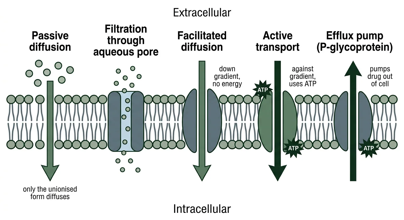
Figure 1 — Mechanisms of drug transport across the cell membrane. Cross-sectional diagram of a cell membrane showing passive diffusion, filtration through aqueous pores, facilitated diffusion, active transport requiring ATP, and P-glycoprotein-mediated efflux pumping drug back out of the cell.
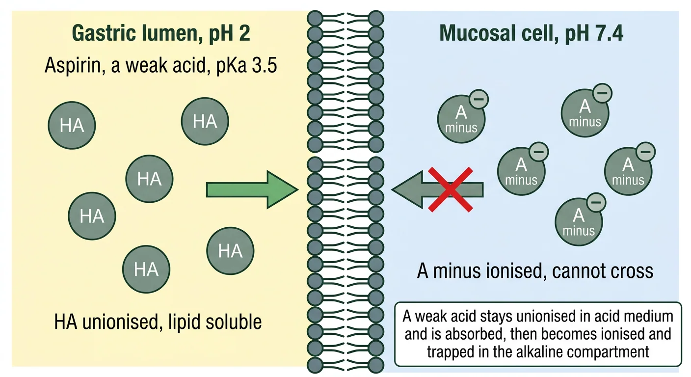
Figure 2 — pH partition and ion trapping across the gastric mucosa. Diagram showing a weak acid remaining unionised in the acidic gastric lumen, diffusing across the membrane into the neutral cell interior where it becomes ionised and can no longer diffuse back, illustrating ion trapping.
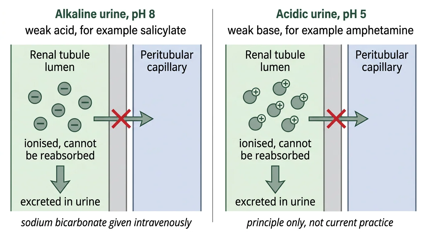
Figure 3 — Urinary ion trapping in poisoning. Two-panel renal tubule diagram showing that alkaline urine ionises and traps weak acids such as salicylate while acidic urine traps weak bases such as amphetamine, in each case preventing tubular reabsorption and enhancing excretion.
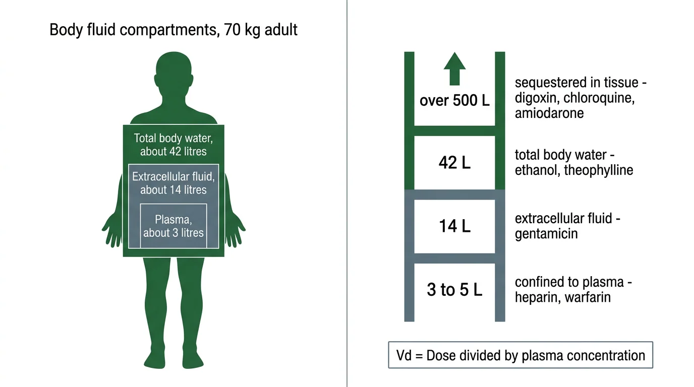
Figure 4 — Apparent volume of distribution and the body fluid compartments. Infographic showing the plasma, extracellular fluid and total body water compartments of a 70 kg adult alongside a scale of volume of distribution values with representative drugs, from heparin confined to plasma to chloroquine extensively sequestered in tissue.
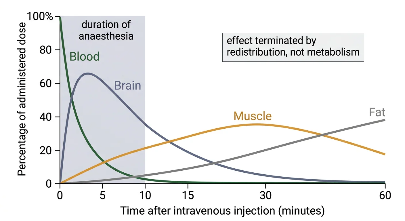
Figure 5 — Redistribution of thiopentone. Graph of thiopentone concentration over time in blood, brain, muscle and fat, showing the rapid rise and fall of brain concentration terminating anaesthesia within about ten minutes as drug redistributes to muscle and then fat.
Clinical Correlation
Vignette 1 — Salicylate overdose and urinary alkalinisation
A 19-year-old woman is brought in four hours after ingesting about 30 tablets of aspirin. She is hyperventilating, complains of tinnitus, and arterial blood gas shows a mixed respiratory alkalosis and metabolic acidosis. Serum salicylate is markedly elevated.
Reasoning: Beyond supportive care and fluid resuscitation, elimination is enhanced by intravenous sodium bicarbonate to raise the urine pH to about 7.5–8.0, with careful potassium replacement (hypokalaemia prevents effective alkalinisation because the kidney will exchange H⁺ for Na⁺ instead of K⁺). The pharmacological basis is ion trapping: salicylate is a weak acid, and in alkaline tubular fluid it exists almost entirely as the ionised salicylate anion, which cannot be passively reabsorbed across the tubular epithelium and is therefore carried out in the urine. Alkalinisation also reduces movement of salicylate into the central nervous system by favouring the ionised form in plasma. Salicylate is additionally a good candidate for haemodialysis in severe poisoning because it has a low volume of distribution, low molecular weight and, at toxic concentrations, saturated protein binding — all the features that make a drug dialysable.
Vignette 2 — Cotrimoxazole in a neonate
A 3-week-old jaundiced neonate is prescribed cotrimoxazole for a presumed infection. The paediatric registrar stops the prescription immediately.
Reasoning: Sulfonamides are highly bound to albumin and compete with bilirubin for the same binding sites. In the neonate, displacement raises the free unconjugated bilirubin concentration at a time when hepatic glucuronyl transferase activity is low and the blood–brain barrier is immature. Free lipid-soluble unconjugated bilirubin crosses into the brain and is deposited in the basal ganglia and brainstem nuclei, producing kernicterus — a permanent choreoathetoid, deafness-associated encephalopathy. Sulfonamides are therefore contraindicated in neonates under about two months of age and in the third trimester of pregnancy. This vignette illustrates the general rule that a displacement interaction becomes dangerous when the displaced substance is highly bound, has a small volume of distribution, and cannot be rapidly eliminated.
Vignette 3 — Why thiopentone wears off, and why the third dose does not
A patient is induced with intravenous thiopentone and wakes after about eight minutes. During a prolonged procedure repeated boluses are given, and recovery afterwards takes several hours.
Reasoning: Thiopentone is extremely lipid-soluble and reaches the richly perfused brain within one circulation time, producing anaesthesia almost immediately. Recovery from a single dose occurs because the drug redistributes down its concentration gradient from the brain into skeletal muscle, and subsequently into fat — tissues that are far less well perfused but far larger in aggregate capacity. The effect is terminated by redistribution, not by metabolism or excretion; the elimination half-life remains many hours and the drug persists in the body long after consciousness returns. With repeated dosing the peripheral tissues become saturated, so no gradient remains to draw drug out of the brain, and termination now depends on the slow process of hepatic metabolism — hence the prolonged recovery, a phenomenon termed cumulation. Patients with a small muscle mass, and the elderly, are correspondingly more sensitive.
Vignette 4 — Digoxin toxicity and the limits of dialysis
A patient with digoxin toxicity and renal failure is receiving haemodialysis. The family asks whether dialysis will remove the drug.
Reasoning: It will not, to any useful extent. Digoxin has a very large volume of distribution (of the order of 500 L) because it binds avidly to Na⁺/K⁺-ATPase in cardiac and skeletal muscle. The overwhelming majority of the drug in the body is therefore in tissue, not in the plasma presented to the dialyser, and removing plasma drug simply allows re-equilibration from the tissue reservoir. The correct treatment for significant toxicity is digoxin-specific antibody fragments (Fab), together with correction of electrolytes. The contrast is instructive: lithium, methanol, ethylene glycol, salicylate and phenobarbitone all have low volumes of distribution and low protein binding, and are removed effectively by dialysis, whereas digoxin, amiodarone, tricyclic antidepressants and benzodiazepines are not.
Practical Linkage
Linked competencies: PH1.4, PH2.4 (calculation of drug dosage) — see also PR02 (Clinical pharmacy exercises).
Worked calculation exercises. Students should complete these with attention to unit conversion, which is the commonest source of error.
Problem 1 — Volume of distribution
A 60 mg intravenous bolus of a drug produces an extrapolated plasma concentration at time zero of 1.5 mg/L. Calculate Vd.
Solution: Vd = Dose / C₀ = 60 mg ÷ 1.5 mg/L = 40 L. This approximates total body water, suggesting a small, relatively lipid-soluble, poorly protein-bound drug.
Problem 2 — Unit-conversion trap
A 500 mg dose gives a plasma concentration of 5 mg/dL. Calculate Vd.
Solution: First convert: 5 mg/dL = 50 mg/L (multiply by 10, since 1 L = 10 dL). Vd = 500 ÷ 50 = 10 L. A candidate who omits the conversion obtains 100 L, a tenfold error.
Problem 3 — Loading dose
A drug has a Vd of 35 L. The target plasma concentration is 4 mg/L. Calculate the intravenous loading dose, and then the oral loading dose if oral bioavailability is 0.5.
Solution: Loading dose = (Target concentration × Vd) / F. Intravenously, F = 1, so LD = 4 × 35 = 140 mg. Orally, LD = 140 ÷ 0.5 = 280 mg.
Problem 4 — Percentage ionisation
A weak acid has a pKa of 4.4. What fraction is ionised in plasma at pH 7.4?
Solution: pH − pKa = 3. For a weak acid, log([ionised]/[unionised]) = 3, so the ratio is 1000:1 — the drug is approximately 99.9% ionised and essentially confined to the extracellular fluid.
Problem 5 — Bioavailability
The same 100 mg dose of a drug produces an AUC of 40 mg·h/L intravenously and 26 mg·h/L orally. Calculate the absolute bioavailability.
Solution: F = AUC(oral)/AUC(IV) = 26 ÷ 40 = 0.65, that is 65%.
Problem 6 — Applied reasoning
A patient has taken an overdose of a drug with a Vd of 800 L and 95% plasma protein binding. Should haemodialysis be offered?
Expected answer: No. The high Vd indicates that almost all the drug is sequestered in tissue, and the high protein binding further restricts the free fraction available for filtration; dialysis will remove a negligible proportion of the body burden.
MCQ Bank
40 questions · tagged by topic, exam pattern & difficulty · full explanations
Which of the following is a characteristic feature of passive diffusion but NOT of carrier-mediated transport?
Rapid Revision
- Predominant mechanism of drug transport across membranes — Passive diffusion of the unionised form
- Three features of carrier-mediated transport — Saturability, structural specificity and competitive inhibition
- Only form of a drug that crosses membranes — The unionised, lipid-soluble form
- Henderson-Hasselbalch for a weak acid — pH equals pKa plus log of ionised over unionised
- Percentage ionised when pH equals pKa — Exactly 50%
- Ionisation when pH and pKa differ by one unit — Approximately 91% to 9%
- Weak acids are better absorbed from — The stomach, though the intestine absorbs more in absolute terms because of surface area
- Site of maximum absorption of nearly all oral drugs — Small intestine, because of its vast surface area
- Urinary alkalinisation is used in poisoning by — Salicylate and phenobarbitone, both weak acids
- Target urine pH in salicylate poisoning — Approximately 7.5 to 8.0
- Why local anaesthesia fails in an abscess — Acidic tissue ionises the weak base, reducing membrane penetration
- Drugs concentrated in breast milk — Weak bases, because milk is slightly acidic relative to plasma
- Formula for absolute bioavailability — AUC oral divided by AUC intravenous for the same dose
- Regulatory bioequivalence limits — 90% confidence interval within 80 to 125 per cent
- Parameter reflecting rate rather than extent of absorption — Tmax, together with Cmax
- Formula for volume of distribution — Dose divided by plasma concentration at time zero
- Approximate plasma, ECF and total body water volumes in a 70 kg adult — 3 litres, 14 litres and 42 litres
- Drug with high lipid solubility but low Vd — Warfarin, because 99% albumin binding predominates
- Implication of a very high volume of distribution — Extensive tissue sequestration and poor removal by dialysis
- Drugs well removed by haemodialysis — Lithium, methanol, ethylene glycol, salicylate and phenobarbitone
- Drugs poorly removed by haemodialysis — Digoxin, amiodarone, tricyclic antidepressants and benzodiazepines
- Loading dose formula — Target concentration multiplied by Vd, divided by bioavailability
- Plasma protein binding acidic drugs — Albumin
- Plasma protein binding basic drugs — Alpha-1-acid glycoprotein, an acute-phase reactant
- Most examined displacement interaction — Sulfonamides displacing bilirubin in the neonate, causing kernicterus
- Conditions for a clinically significant displacement interaction — High binding above 90%, narrow therapeutic index and small volume of distribution
- Efflux pump limiting brain and fetal drug entry — P-glycoprotein
- Opioid with no central effect owing to P-glycoprotein efflux — Loperamide
- Anticoagulant of choice in pregnancy — Heparin, because it does not cross the placenta
- Cause of the brief action of thiopentone — Redistribution from brain to muscle and fat
- Reason recovery is prolonged after repeated thiopentone doses — Saturation of peripheral tissues abolishes the redistribution gradient
- Brain region lacking a blood-brain barrier — The chemoreceptor trigger zone in the area postrema
Viva Questions
- Define pharmacokinetics — The study of the absorption, distribution, metabolism and excretion of drugs, that is what the body does to the drug.
- What is the pH partition hypothesis — Only the unionised lipid-soluble form of a weak electrolyte crosses a biological membrane by passive diffusion, so the pH of the compartment relative to the drug pKa determines its distribution.
- Define pKa — The pH at which a drug is exactly 50% ionised and 50% unionised.
- What is ion trapping — Accumulation of a drug on one side of a membrane because the pH there converts it to the ionised non-diffusible form.
- How would you enhance elimination of salicylate in overdose — Alkalinise the urine with intravenous sodium bicarbonate to a pH of about 7.5 to 8.0, with potassium replacement, and consider haemodialysis in severe cases.
- Define bioavailability and state how it is measured — The fraction of an administered dose reaching the systemic circulation unchanged, measured as the ratio of the AUC by the test route to the AUC after intravenous administration of the same dose.
- Differentiate bioequivalence from therapeutic equivalence — Bioequivalence is similarity of the plasma concentration-time profile, whereas therapeutic equivalence is equivalence of clinical effect and safety, for which bioequivalence is accepted as a surrogate.
- Define apparent volume of distribution and explain why it is called apparent — It is the hypothetical volume of fluid needed to contain the total body drug at the measured plasma concentration; it is apparent because it is a calculated proportionality constant and may greatly exceed any real body volume.
- Why does warfarin have a low volume of distribution despite being lipid soluble — Because it is 99% bound to plasma albumin and bound drug cannot leave the vascular compartment, so protein binding predominates over lipid solubility.
- Which drugs are removed effectively by dialysis and why — Those with a low volume of distribution, low plasma protein binding, low molecular weight and water solubility, such as lithium, methanol and salicylate.
- Name the two principal drug-binding plasma proteins and their substrates — Albumin for acidic and neutral drugs, alpha-1-acid glycoprotein for basic drugs.
- When is a protein-binding displacement interaction clinically important — When the displaced drug is more than 90% bound, has a narrow therapeutic index and a small volume of distribution, as with warfarin and phenytoin.
- Why are sulfonamides avoided in neonates — They displace bilirubin from albumin, and free bilirubin crosses the immature blood-brain barrier to cause kernicterus.
- What terminates the action of a single dose of thiopentone — Redistribution from the brain to skeletal muscle and then fat, not metabolism.
- What is P-glycoprotein and why does it matter — An ATP-dependent efflux pump in the gut, biliary canaliculus, renal tubule, placenta and blood-brain barrier that extrudes drugs, reducing oral bioavailability and limiting entry into the brain and fetus.
- Which anticoagulant is safe in pregnancy and why — Heparin, because it is large and highly polar and does not cross the placenta.
References
- Tripathi KD. Essentials of Medical Pharmacology. 9th ed. New Delhi: Jaypee Brothers Medical Publishers; Chapters 2 and 3 (Pharmacokinetics: Membrane transport, absorption and distribution of drugs).
- Katzung BG, Vanderah TW (eds). Basic & Clinical Pharmacology. 16th ed. New York: McGraw Hill; Chapter 1 (Introduction) and Chapter 3 (Pharmacokinetics & Pharmacodynamics: Rational Dosing).
- Brunton LL, Knollmann BC (eds). Goodman & Gilman's The Pharmacological Basis of Therapeutics. 14th ed. New York: McGraw Hill; Chapter 2 (Pharmacokinetics: The Dynamics of Drug Absorption, Distribution, Metabolism and Elimination).
- Ritter JM, Flower RJ, Henderson G, et al. Rang & Dale's Pharmacology. 10th ed. Edinburgh: Elsevier; Chapters 9 and 10 (Absorption and Distribution of Drugs; Pharmacokinetics).
- National Medical Commission. Competency Based Undergraduate Curriculum for the Indian Medical Graduate, Volume II — Pharmacology (Code: PH), competency PH1.4.
- Shargel L, Yu ABC. Applied Biopharmaceutics and Pharmacokinetics. 7th ed. New York: McGraw Hill; chapters on drug absorption, bioavailability and bioequivalence.
- Proudfoot AT, Krenzelok EP, Vale JA. Position paper on urine alkalinization. Journal of Toxicology: Clinical Toxicology 2004;42(1):1–26.
- Holford NHG. Volume of distribution. Translational and Clinical Pharmacology 2016;24(2):74–77.
Downloads
All deliverables & printable revision sheets for CH02
Printable Chapter Handout & Study Notes
Generate a clean, high-yield PDF or print directly without sidebars and navigation menus.